We present the solution structures of HIV-1 proteins NC(p7)1-55 corresponding to the full-length NC(p7) and mature p6. The studies were carried in water and, to mimic the membrane, in micellar DPC (Dodecylphosphocholine) conditions. Our results unravel for the first time the structure adopted by the N-terminal amino acids of the free NC(p7)1-55, with the formation of a small helix spanning residues F6 to R10. Our NMR and Fluorescence Anisotropy data disclose an interaction between NC(p7)1-55 and p6 both in water and DPC, with respective Kd of 2.5mM and 370 mM at 23°C. The interaction is thus strengthened in lipidic conditions. Protein p6 stabilizes the N-terminus of NC(p7)1-55 while increasing at the same time the dynamic of the first zinc finger. Although the entire p6 sequence is involved in the interaction, we show that its C-terminal region is particularly sensitive to the presence of NC(p7)1-55, with a propensity of forming a a helix ranging from amino acids S111 to F116. This study brings experimental evidence of a direct protein-protein interaction between p6 and the N-terminal region of NC(p7)1-55. We further show that such interaction is readily accommodated within the NC(p15) framework and hypothesize that it may facilitate the selective assembly of assembly of the viral genomic RNA (gRNA) in the cell.
Supplementary Table 1. Clinical metadata Supplementary Table 2. 3D stacked serial sections overview Supplementary Table 3. 2D gene discovery sections overview Supplementary Table 4.CODEX panel information Supplementary Table 5. Xenium panel information Supplementary Table 6. HT704B1 distant region path metadata Supplementary Table 7: Section retention for 3D cohort Supplementary Table 8. Model architecture, hyperparameters, and training details for H&E protein prediction Supplementary Table 9. 3D serial section ROI summary metadata Supplementary Table 10. 3D serial section ROI path metadata Supplementary Table 11. 2D marker gene discovery: section-level feature metrics Supplementary Table 12. 2D marker gene discovery: visium barcode annotations Supplementary Table 13. 3D ROI manual marker gene discovery: feature expression for annotated regions Supplementary Table 14. 3D ROI manual marker gene discovery: feature fold change for annotated regions Supplementary Table 15: Data associated with breast functional assays Supplementary Table 16: Data associated with prostate functional assays.
Erik Storrs, Chia-Kuei Mo, Wen-hung Chou et al.· 0 citations
Chromosome conformation capture methods such as Hi-C and Micro-C have transformed our understanding of genome organisation, revealing architectural features from megabase-scale compartments to kilobase-scale loops. Yet the choice of fragmentation enzyme imposes constraints that shape which features are most readily observed. Restriction enzyme workflows rely on sequence specific cut sites and often use detergent conditions that can perturb chromatin structure, while micrococcal nuclease (MNase) exhibits pronounced sequence specificity and generates termini that require end processing before efficient ligation. These properties add time, introduce variability, and may bias fragment recovery. This thesis develops CAD-C, a chromosome conformation capture method that exploits the apoptotic nuclease CAD (Caspase-Activated DNase) to address these limitations. CAD is a double strand-specific endonuclease that cleaves preferentially in linker DNA between nucleosomes and generates predominantly blunt or near-blunt double-stranded breaks with 5′-phosphate and 3′ hydroxyl termini, the end chemistry preferred by T4 DNA ligase. This native ligation competence can eliminate enzymatic end-repair steps in Direct CAD-C and supports efficient proximity ligation under chromatin-preserving conditions. I first describe the design, expression, and purification of a recombinant CAD construct optimised for chromatin fragmentation. Because CAD requires its inhibitor-chaperone ICAD for proper folding, I developed a dual-cassette bacterial expression system that co-expresses CAD with a TEV cleavable ICAD variant, enabling on-demand activation of the nuclease. I also identify purification conditions that reduce contaminating bacterial nuclease activity and improve preparation quality for controlled chromatin digestion. I next characterise CAD cleavage patterns on crosslinked chromatin and introduce the CHOMP principle (CAD Hindered Overdigestion at Margins of Proteins), which describes how CAD’s steric bulk can bias cleavage toward defined offsets from chromatin-bound proteins, leaving accessible termini available for ligation. This framework provides a conceptual basis for interpreting CAD-C contact maps and the locations where ligation-competent ends form. I then present the CAD-C protocol in three workflow variants: Classic, Express, and Direct. Benchmarking against Micro-C and Hi-C in GM12878, hTERT-RPE1, and K562 cells shows that CAD-C recovers major architectural features, including compartments, domains, and loops, with performance comparable to Micro-C in these datasets. At the same time, CAD-C exhibits a distinctive short-range contact signature and preferentially recovers a subset of active regulatory contacts, including promoter-associated loops. I develop a ligation-gating model to explain how CAD’s native ligation competence can increase near-diagonal contact density while sharpening loop contrast relative to MNase-based methods. Finally, I introduce CADwalks, a per-molecule analytical framework for Direct CAD-C con catemers. Because CAD generates nucleosome-resolution fragments whose boundaries encode protein-DNA footprint information, and because its ligation-ready termini support efficient concate mer formation, CADwalks preserve the relative order, orientation, and co-occurrence of fragments captured on the same concatemer molecule, information that pairwise decomposition discards. At CTCF boundaries, CADwalks reveal a nucleosome-scale directional asymmetry consistent with oriented loop extrusion. This thesis is primarily a tool-development and validation effort. The experimental work em phasizes proof-of-principle demonstrations rather than exhaustive perturbation biology. Together, CAD-C and CADwalks show that changing the chromatin fragmentation enzyme changes not only assay resolution and workflow efficiency, but also the biochemical gate through which contacts are captured. They establish a nucleosome-resolution, per-molecule framework for chromosome con formation capture and provide a foundation for future studies of chromatin architecture, regulatory looping, and gene control.
Platelet-erythrocyte (PE) complexes, composed of single erythrocytes bound to platelets, are a physiological component of the bloodstream and contribute to erythrocyte turnover. Conditions in which PE complex production is increased or their clearance from the circulation is impeded are associated with hypercoagulable states and thrombocytopenia. Sepsis is a serious acute inflammatory condition that is often complicated by major hemostatic derangements, including thrombosis, anemia and thrombocytopenia, and an important driver of sepsis pathology are extracellular histones, which are released into the circulation from activated neutrophils. Histone-neutralizing small polyanions (SPAs) have been developed to block the cytotoxic effects of histones and are beneficial in treating sepsis. This study investigated whether histones may stimulate increased PE complex production as a potential cause of the hemostatic complications in disease, and whether SPAs can prevent complex generation. Treatment with histones in co-incubated human erythrocytes and platelets, or murine blood, increased PE complex production approximately 10-fold compared with control conditions. Mice administered histones by intravenous injection displayed reduced platelet counts but sustained complex levels, suggesting complex production was enhanced within the diminished platelet pool. The platelets in the histone-stimulated complexes were also more activated than cell-free platelets, according to increased surface expression of P-selectin and the fibrin-binding form of integrin αIIbβ3, determined using flow cytometry, and their altered shape and presence of filopodia observed by scanning electron microscopy. Inclusion of SPAs in the histone-treated cells and blood prevented histone-stimulated production of complexes, indicating the importance of the histone-mediated cell injury in their generation and identifying PE complexes as a treatment target. Why was the study done? Platelets can attach to red blood cells to form platelet–erythrocyte (PE) complexes, which normally help remove old or damaged red blood cells. However, excessive complex formation may contribute to blood clotting disorders and low platelet counts in diseases like sepsis. In these conditions, proteins called histones are released into the bloodstream and can damage cells. This study investigated whether histones, as a result of damaging erythrocytes, increase PE complex formation and whether drugs that neutralize histones can prevent this. What did the researchers do? Researchers examined human blood cells in the laboratory and mouse blood samples to measure PE complex formation after exposure to histones. They also assessed platelet activation and tested two histone-neutralizing compounds (small polyanions, SPAs) to see if they could block these effects. Additional experiments were conducted in mice given histones. What did the researchers find? Histones increased PE complex formation by about 10-fold and made platelets more activated in the blood cells and samples. In live mice, histone administration sustained complex formation despite the histones also reducing the number of available platelets. The SPA compounds prevented complex formation when added before or alongside histones but were ineffective after complexes had formed. What do the findings mean? Histones may drive harmful clotting and lead to low platelet counts by increasing PE complexes and platelet activation. Drugs that neutralize histones could help prevent these effects and represent a potential treatment approach.
Platelet-erythrocyte (PE) complexes, composed of single erythrocytes bound to platelets, are a physiological component of the bloodstream and contribute to erythrocyte turnover. Conditions in which PE complex production is increased or their clearance from the circulation is impeded are associated with hypercoagulable states and thrombocytopenia. Sepsis is a serious acute inflammatory condition that is often complicated by major hemostatic derangements, including thrombosis, anemia and thrombocytopenia, and an important driver of sepsis pathology are extracellular histones, which are released into the circulation from activated neutrophils. Histone-neutralizing small polyanions (SPAs) have been developed to block the cytotoxic effects of histones and are beneficial in treating sepsis. This study investigated whether histones may stimulate increased PE complex production as a potential cause of the hemostatic complications in disease, and whether SPAs can prevent complex generation. Treatment with histones in co-incubated human erythrocytes and platelets, or murine blood, increased PE complex production approximately 10-fold compared with control conditions. Mice administered histones by intravenous injection displayed reduced platelet counts but sustained complex levels, suggesting complex production was enhanced within the diminished platelet pool. The platelets in the histone-stimulated complexes were also more activated than cell-free platelets, according to increased surface expression of P-selectin and the fibrin-binding form of integrin αIIbβ3, determined using flow cytometry, and their altered shape and presence of filopodia observed by scanning electron microscopy. Inclusion of SPAs in the histone-treated cells and blood prevented histone-stimulated production of complexes, indicating the importance of the histone-mediated cell injury in their generation and identifying PE complexes as a treatment target. Why was the study done? Platelets can attach to red blood cells to form platelet–erythrocyte (PE) complexes, which normally help remove old or damaged red blood cells. However, excessive complex formation may contribute to blood clotting disorders and low platelet counts in diseases like sepsis. In these conditions, proteins called histones are released into the bloodstream and can damage cells. This study investigated whether histones, as a result of damaging erythrocytes, increase PE complex formation and whether drugs that neutralize histones can prevent this. What did the researchers do? Researchers examined human blood cells in the laboratory and mouse blood samples to measure PE complex formation after exposure to histones. They also assessed platelet activation and tested two histone-neutralizing compounds (small polyanions, SPAs) to see if they could block these effects. Additional experiments were conducted in mice given histones. What did the researchers find? Histones increased PE complex formation by about 10-fold and made platelets more activated in the blood cells and samples. In live mice, histone administration sustained complex formation despite the histones also reducing the number of available platelets. The SPA compounds prevented complex formation when added before or alongside histones but were ineffective after complexes had formed. What do the findings mean? Histones may drive harmful clotting and lead to low platelet counts by increasing PE complexes and platelet activation. Drugs that neutralize histones could help prevent these effects and represent a potential treatment approach.
Extracellular RNAs are found in the plant extracellular space, but how they are exported from cells remains unclear. We found that the plant vacuole is a major source of extracellular RNA and identified a class of large extracellular vacuole-derived bodies, which we termed EVacs, that are key mediators of this transport. EVacs are marked by the vacuolar membrane (tonoplast) proteins γ-TIP and V-ATPase and originate as intravacuolar structures formed by inward folding of the tonoplast, encapsulating intact cytoplasmic material, including both RNAs and proteins. These intravacuolar bodies then escape the vacuole and are subsequently released from the plasma membrane of mesophyll cells into the apoplast. These findings provide a novel mechanism for the unconventional secretion of macromolecules in plants.
Lucía Borniego, Meenu Singla‐Rastogi, Megha Hastantram Sampangi-Ramaiah et al.· bioRxiv (Cold Spring Harbor...· 0 citations
Single-cell proteomics (SCP) has emerged as a powerful approach to quantify protein expression variability at cellular resolution, yet most state-of-the-art workflows are tailored to eukaryotic cells with only one study exploring how single bacteria can be analyzed by mass spectrometry. Here, we established bacSCP, a protocol extending SCP to bacterial cells, facing analytical challenges such as the thick bacterial cell wall hampering lysis, the extremely small cell size and resultant low protein content, and the comparatively high level of contaminating proteins from external sources. Using this bacSCP pipeline, we quantified more than 50 bacterial proteins from single
Bacillus subtilis
and
Escherichia coli
cells. Upon heat stress, we reproducibly observed up to 8-fold upregulation of chaperones including GroEL, GroES, and ClpC for a
B. subtilis ΔmcsB
strain. Importantly, single-cell measurements revealed potential heterogeneity within the heat-stressed subpopulation, enabling interrogation of stress-response variability at the proteome level. These results demonstrate the feasibility of bacSCP and provide a foundation for studying bacterial stress adaptation and phenotypic diversity with single-cell proteomic resolution.
Julia Leodolter, Tim Thierer, K. Mechtler et al.· Nature Communications· 0 citations
OBJECTIVES
Acute pancreatitis (AP) is a painful and potentially life-threatening disorder, with no effective therapy available to date. Genome assessment shows promise as a potential method for predicting AP severity. We hypothesized that single nucleotide polymorphisms (SNPs) and expression changes in Heat Shock Protein 70 (HSP70) family genes may play a role in early prediction of AP severity.
METHODS
A total of 44 AP patients and 47 age- and sex-matched healthy controls were studied. Leukocyte-extracted total RNA and genomic DNA were used for HSPA1A and HSPA1L gene expression and genotyping in promoter regions (HSPA1A: rs1008438 and HSPA1L: rs2227956) analyses, respectively.
RESULTS
No statistically significant differences were found between the genotypes of AP patients and controls, however, HSPA1A expression was 2-fold lower in AP. Decrease in expression was more evident in mild and mild/moderate AP groups vs. controls (P=0.024 and P=0.008, respectively). HSPA1L expression in AG vs. AA carriers was higher in entire cohort (P=0.014) and markedly differed between AP patients (P=0.001). Also, HSPA1L expression was the highest in severe AG cases and significantly differed from AA genotype carriers (P=0.005).
CONCLUSIONS
We suggest that HSP70 family genes expression may be a valuable marker for predicting disease severity in AP.
A. Kielaite-Gulla, J. Gaiževska, A. Šeštokaitė et al.· Pancreas· 0 citations
This manuscript proposes a theoretical application of the General Epistemic Dispersion Theory (GEDT) to the domain of molecular biology, utilizing adult human hemoglobin (HbA) as a model study. The work formalizes protein folding as a configurational reconstruction problem, investigating how informational load is partitioned between structural constraints and dynamic assistance. The study introduces a conditional decomposition of configurational uncertainty, H(Π | F) = I(Π ; S | F) + H(Π | F, S), mapping the primary amino acid sequence to the structural core (S) and the physicochemical environment to the finite space of feasible configurations (𝒫). Through the comparative analysis of HbA assembly, the role of the Alpha-Hemoglobin Stabilizing Protein (AHSP), and specific variants (HbS and Hb G-Copenhagen), the paper examines the informational boundaries of spontaneous folding and dynamic chaperone intervention. This preprint does not introduce novel biochemical mechanisms nor present new experimental biological data.The primary objective is to establish a falsifiable and operationalizable informational framework to quantify the residual configurational uncertainty in biological systems, offering a purely epistemic perspective on the structural economy of molecular folding.
Massimo Comitato· Zenodo (CERN European Organi...· 0 citations
Type 2 diabetes mellitus (T2DM) remains a global health crisis, with nearly 589 million adults currently affected and projections pointing toward 853 million by 2050. Despite an expanding pharmacological armamentarium, a significant proportion of patients fail to achieve adequate glycaemic control, and the limitations of existing therapies, including adverse effects, cost, and limited accessibility, underscore the compelling need for novel therapeutic approaches. Phytochemicals such as curcumin, berberine, quercetin, resveratrol, and epigallocatechin gallate possess well-documented antidiabetic activity, operating through the PI3K/Akt, AMPK (activated protein kinase), NF-κB/JNK (nuclear factor kappa-B), and GLP-1R (glucagon-like peptide-1) signalling axes to improve insulin sensitivity, suppress gluconeogenesis, protect pancreatic beta-cells, and attenuate chronic metabolic inflammation. However, their clinical utility has been fundamentally constrained by poor oral bioavailability arising from low aqueous solubility, gastrointestinal instability, extensive first-pass metabolism, and P-glycoprotein-mediated efflux. Advanced drug delivery systems, including liposomes, solid lipid nanoparticles (SLN), nanostructured lipid carriers, PLGA (Poly (lactic-co-glycolic acid)) and chitosan nanoparticles, nanoemulsions, self-nanoemulsifying drug delivery systems, and phytosomes have demonstrated the capacity to overcome these barriers, achieving five- to ten-fold improvements in systemic bioavailability and substantially enhanced antidiabetic efficacy in preclinical models. Emerging mechanistic evidence further positions gut microbiota modulation and epigenetic reprogramming as additional therapeutic axes through which nano-encapsulated phytochemicals may exert durable metabolic benefits. Nonetheless, critical translational challenges persist, encompassing nanotoxicological risks, herb–drug interactions, the absence of harmonised regulatory frameworks for nano-phytomedicine products, phytochemical raw material variability, and formidable technical and economic barriers to scalable nanoparticle manufacturing. This review synthesises the current mechanistic, formulation, and clinical evidence within a unified analytical framework and identifies the strategic priorities of rigorous clinical development, regulatory clarity, and manufacturing standardisation required to translate nano-phytomedicine science into evidence-based T2DM therapeutics.
A. Dadhich, Vikas Sharma, Shivika Sharma et al.· Pharmaceutics· 0 citations
PRMT5 and RBM39 inhibition regulate DNA repair and metabolic pathways. A, Workflow of the RNA-seq approach: PSN1 cells were treated with DMSO as the vehicle control, 4 nmol/L JNJ-64619178, 200 nmol/L indisulam, or the combination of 4 nmol/L JNJ-64619178 and 200 nmol/L indisulam for 48 hours, mRNA was isolated, and mRNA sequencing with subsequent GSEA was performed. The experiment was conducted as four biological replicates. B, Venn diagram showing downregulated GO-BP gene sets in PSN1 cells; all treatments were compared with the vehicle control in individual GSEAs. Only gene sets with an FDR < 0.05 were considered significant and used for the analysis. C, 24 GO-BP gene sets downregulated exclusively in the combination treatment are indicated. The value of the normalized enrichment score (NES) is color-coded. D, Number and composition of significant changed alternative splicing (AS) events observed upon monotherapy or combination treatment of JNJ-64619178 and/or indisulam. E, Percent spliced-in (ΔPSI) distribution of different AS events identified following monotherapy or combination treatment with JNJ-64619178 and/or indisulam. P values based on the Wilcoxon test are indicated. F, PSN1 cells were either treated with DMSO control or 4 nmol/L JNJ-64619178 and 200 nmol/L indisulam for 96 hours followed by mass spectrometry determination of the proteome. G, Volcano plot of deregulated proteins in PSN1 cells treated with the combination therapy [see (F)] vs. control. Proteins with an adjusted P value < 0.05 were colored. Proteins involved in the DNA repair pathway are highlighted lilac, whereas proteins involved in the splicing pathway are highlighted pink. H, ΔPSI distribution of different AS events in the DNA damage signaling and repair pathway genes ATM, FAN1, and FANCL observed upon monotherapy or the combination treatment with JNJ-64619178 and/or indisulam. I, Results of the γH2A.X phosphorylation assay: PSN1 cells were treated with 50 nmol/L MRTX1719 and/or 240 nmol/L indisulam for 48 hours (left) or 72 hours (right). For negative control, cells were left vehicle-treated; for positive control, cells were irradiated with 2 Gy for 30 minutes. The fold increase of γH2A.X phosphorylation is shown on the y-axis. Statistical analysis was performed using a repeated-measures one-way ANOVA with correction for multiple-testing according to the Tukey test: **, P < 0.01; *, P < 0.05, n = 3. A3SS, alternative 3′ splice site; A5SS, alternative 5′ splice site; MSE, mutually exclusive exon; RI, retained intron; SI, skipped exon. [A, Created in BioRender. Schneider, G. (2026) https://BioRender.com/fwl2ir3; F, Created in BioRender. Schneider, G. (2026) https://BioRender.com/z84e701.]
Valentina Spielmann, Jonas Buchloh, Selen Selcen et al.· 0 citations
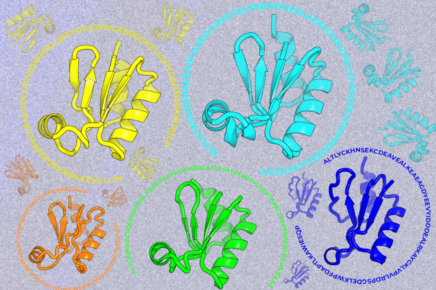
A new machine-learning framework aims to improve the success rate of computational protein design while moving away from results that reproduce sequences found in nature.