Data indicate that platelets support greater activity of FVIII than PLV through stabilization against dissociation of the A2 domain and protection from degradation by APC.
Abstract
Activated platelets support factor VIII (FVIII) activity at 100-fold lower FVIII concentration than phospholipid vesicles (PLV) but the mechanism(s) remain unknown. Therefore, we compared activity of FVIIIa vs. emicizumab using platelets and PLV. We also utilized engineered B-domain deleted human factor VIII (FVIIIBDD) with degradation-resistant primary APC cleavage sites R336Q/R562Q (FVIIIQQ) or a stabilized A2 domain, D519V/E665V (FVIIIVV), or both (FVIIIQQVV). FVIII-dependent clotting was faster with platelets than PLV. The relative platelet:PLV FVIII procoagulant activity varied from 40:1 to 2:1; the highest ratios achieved with lower FVIII concentrations. In contrast, activity of Emi was similar with platelets and PLV. Steady state kinetic experiments indicated that platelets do not convey superior Xase activity. This indicated that FVIII binding sites protect or stabilize FVIII(a). FVIIIVV and FVIIIQQVV supported equivalent clot times on platelets and PLV suggesting that platelets slow dissociation of the A2 subunit. Studies with purified proteins confirmed that FVIIIa dissociates more slowly on platelets than on PLV. Addition of APC prevented PLV-supported clotting but modestly slowed clotting with FVIIIQQ and PLV or with FVIIIBDD and platelets. This suggested that platelets protect FVIII from APC. Defined studies confirmed that platelets slowed APC degradation of FVIII(a) to about the same extent as inactivation of FVIIIQQ with PLV. Platelet microparticles slowed FVIII degradation to a degree that was intermediate between PLV and platelets. Together, these data indicate that platelets support greater activity of FVIII than PLV through stabilization against dissociation of the A2 domain and protection from degradation by APC.
FV is an endogenous anticoagulant that inhibits TF-initiated coagulation by limiting FX activation by TF:FVIIa through a membrane-dependent mechanism, which refines models of coagulation initiation and may help explain how FV variation contributes to bleeding and thrombosis.
M. Jewell, Christine H Baird, D. Thornhill et al.· Blood· 0 citations
Factor VIII Aurora (FVIII-R571S) is the first described naturally occurring enhanced-potency FVIII variant identified in a patient with recurrent thrombosis and early mortality. The patient's plasma exhibited increased procoagulant activity and reduced responsiveness to activated protein C (APC). To define the mechanism, we generated recombinant FVIII-R571S and performed in vitro and in vivo studies. Consistent with the clinical phenotype, FVIII-R571S demonstrated a 6-fold increase in one-stage assay activity, while chromogenic substrate assay activity was comparable to wild-type FVIII (FVIII-WT). This discrepancy was explained by biochemical studies showing that activated FVIII-R571S (FVIIIa-R571S) has 10-20-fold higher affinity for FIXa; notably, the chromogenic assay is insensitive to differences in FVIIIa-FIXa affinity. Additional analyses demonstrated that FVIII-R571S is inactivated by APC and protein S analogous to FVIII-WT, indicating that the variant is not intrinsically APC-resistant. However, the increased affinity of FVIIIa-R571S for FIXa confers FIXa-dependent reduced A2-domain dissociation and APC-mediated inactivation in purified and plasma-based studies. These enhanced biochemical properties translated in vivo to a more potent procoagulant phenotype in hemophilia A mice. In the tail clip assay, FVIII-R571S exhibited a 4-5-fold increase in potency compared to FVIII-WT. In a thrombosis model, FVIII-R571S promoted significantly increased platelet and fibrin accumulation relative to FVIII-WT at equivalent antigen levels. Collectively, these data demonstrate that the prothrombotic phenotype of FVIII-R571S is driven by increased FIXa affinity, which enhances FVIIIa-FIXa complex assembly and function. This same mechanism confers reduced A2 dissociation and functional APC resistance, providing a unifying explanation for the observed gain-of-function phenotype.
Johnathan J Morris, Robert J. Davidson, Connor T Watson et al.· Blood· 0 citations
INTRODUCTION
Various extended half-life recombinant factor VIII (EHL-FVIII) products have been designed to improve the pharmacokinetic properties of FVIII, allowing prolonged haemostatic coverage and reducing the injection burden in people with haemophilia A. Nevertheless, the influence of direct molecular attachment on fibrin clot formation and stability remains to be investigated.
AIM
To investigate the stability and architecture of fibrin clots in the presence of various types of EHL-FVIII.
METHODS
Three EHL-FVIII products with direct attachment modifications (damoctocog alfa pegol, efraloctocog alfa, and rurioctocog alfa pegol) and two standard FVIII (turoctocog alfa and rurioctocog alfa) were added to FVIII-deficient whole blood and plasma at various concentrations for functional comparison. Under whole-blood conditions, functional assays were performed using rotational thromboelastometry (ROTEM) and a microchip flow-chamber system (T-TAS). Under plasma-based conditions, fibrin fibres were directly observed by electron microscopy and coagulation function was assessed using clot waveform analysis (CWA). Anticoagulant and fibrinolytic activities were evaluated by CWA with the addition of activated protein C and tissue plasminogen activator, respectively.
RESULTS
At equivalent activity levels, none of the assays revealed significant differences among the three EHL products or the two standard products. All contributed comparably to fibrin clot formation and stability, as well as to anticoagulation and fibrinolysis functions.
CONCLUSION
Direct modification by PEGylation or IgG-Fc fusion to impart EHL characteristics preserves the functional properties of native FVIII.
PLAIN LANGUAGE SUMMARY
People with haemophilia A require treatment with factor VIII (FVIII) to prevent or control bleeding. Some FVIII products are designed to remain active in the body for a longer time, which can reduce the number of injections needed. This extended half-life is achieved by chemically or biologically modifying FVIII, for example by attaching polyethylene glycol (PEG) or the Fc portion of immunoglobulin G. These treatments are known as extended half-life FVIII (EHL-FVIII) products. However, it has not been fully established whether these modifications affect how blood clots form and remain stable. In this study, we compared three EHL-FVIII products with two standard FVIII products using FVIII-deficient blood and plasma. We evaluated clot formation and stability using several laboratory techniques, including whole-blood assays and scanning electron microscopy. We also examined potential differences in anticoagulant and fibrinolytic properties. At comparable FVIII activity levels, we observed no major differences between the EHL-FVIII products and the standard FVIII products in any of the assays performed. These findings suggest that PEGylation or Fc fusion, which are used to extend the half-life of FVIII, do not impair its functional properties related to fibrin clot formation and stability.
N. Shimonishi, K. Nogami· Haemophilia· 0 citations
Targeted modification of regions surrounding the FIXa active site enables allosteric activation, providing a potential foundation for novel bypassing strategies in hemophilia A therapy.
V. Strijbis, K. Cheung, D. Gobbo et al.· Thrombosis Research· 0 citations
The standardized TF/FXIa dual-activated TGA represents a tool for assessing individual coagulation potential in hemophilia A and showed substantial variation in interindividual TG levels among patients with comparable FVIII activity levels.
T. W. van de Berg, Alexandra C. A. Heinzmann, S. Thomassen et al.· TH Open· 0 citations
BACKGROUND
Factor XI (FXI) occupies a unique and clinically significant niche, bridging the tissue factor-driven and contact-driven coagulation pathways. Irrespective of the trigger, activated factor XI (FXIa) contributes to clotting by activating factor IX (FIX). Biochemical studies established that this reaction requires the membrane-binding FIX-Gla domain to engage an exosite on the FXIa Apple 3 (A3) domain, that only become available upon FXI activation. Structural data for FXIa, FIX, FIXaβ, and the FXIa:FIX complex are lacking; current understanding relies on zymogen FXI crystal structures and homology modeling of FXIa after kallikrein.
OBJECTIVES
Elucidate the high-resolution structure of FXIa in functionally relevant conformation in complex with FIX.
METHODS
We utilized cryogenic electron microscopy (cryo-EM) to determine the structures of human FXIa in complex with its full-length substrate, FIX, and activated product, FIXaβ.
RESULTS
We report the first cryo-EM structures of FXIa in complex with its substrate, FIX, and activated FIX (FIXaβ). The structures capture a functionally relevant conformational change in the FXIa catalytic domain and reveals the first view of the entire FIX and FIXaβ. Critically, we visualize the FIX-Gla domain precisely docked to the FXIa-A3 exosite on both subunits of the FXIa dimer. We also define the first step of proteolysis, visualizing the FIX Arg145 inserted into the primary specificity pocket of FXIa.
CONCLUSIONS
The structures define the full FXIa:FIX interface providing a structural template for understanding the sequential activation of FIX and for developing a new class of selective allosteric antithrombotic agents.
Bassem M Mohammed, Samantha Deavila, Tristan Friet et al.· Journal of Thrombosis and Ha...· 0 citations
Related blog posts
MIT News · Artificial Intelligence· news.mit.eduAug 27, 2026
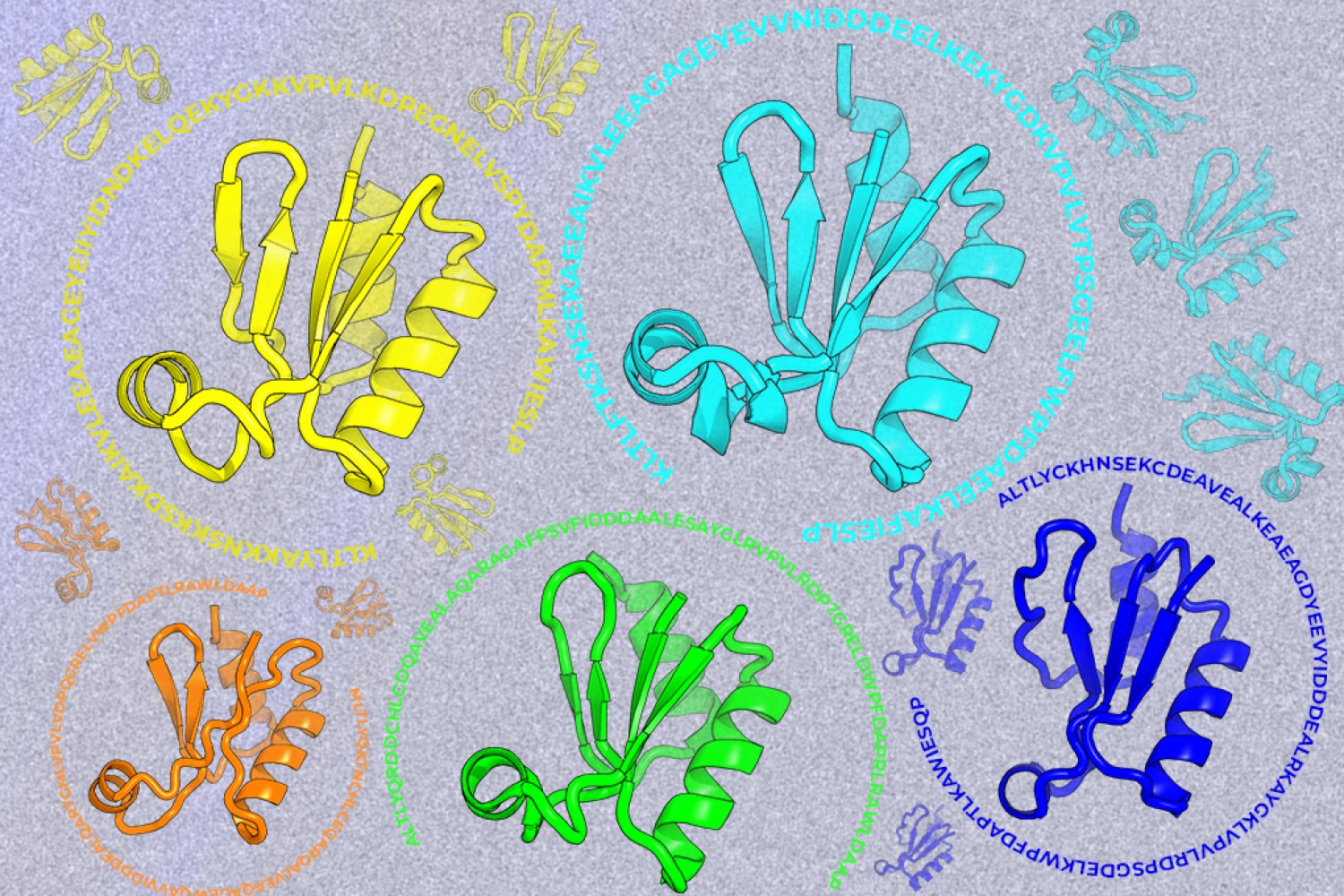
A new machine-learning framework aims to improve the success rate of computational protein design while moving away from results that reproduce sequences found in nature.