Aug 2026· The Journal of the American Dental Association (1939)· 0 citations· 14 references
Medicine
TL;DR
This case illustrates an unusual manifestation of severe jaw pain of unknown origin that was determined to be caused by VOC of the mandible, highlighting the need for dentists to understand SCA pathophysiology and their integral role in the diagnosis of atypical facial pain in patients with SCA.
Abstract
BACKGROUND
Sickle cell anemia (SCA) is an autosomal recessive disorder characterized by a point mutation in the β-globin chain of the hemoglobin protein, causing abnormal folding. This can lead to sickling of red blood cells, agglutination, and ultimately vascular obstruction, which rarely occurs in the cervicofacial region.
CASE DESCRIPTION
This report reviews a rare case of vaso-occlusive crisis (VOC) in the cervical vertebrae and mandible, manifesting as severe mandibular pain of unknown origin in a patient with SCA. The patient experienced diffuse body pain, 3 days in duration, and most prominent in the right hemimandible. Symptoms were refractory to hydration and analgesics, requiring intensive care unit admission. Jaw pain persisted, and magnetic resonance imaging of the face was significant for enhancement in the right posterior hemimandible with surrounding soft-tissue inflammatory changes. Differential diagnoses included osteonecrosis, osteomyelitis, odontogenic infection, extramedullary hematopoiesis, and VOC. The patient lacked signs and sequelae of infection or hematopoiesis; therefore, the pain was attributed to bone infarcts secondary to VOC. The patient was placed on a multimodal analgesic regimen with resolution of symptoms and was discharged in stable condition.
PRACTICAL IMPLICATIONS
This case illustrates an unusual manifestation of severe jaw pain of unknown origin that was determined to be caused by VOC of the mandible. This highlights the need for dentists to understand SCA pathophysiology and their integral role in the diagnosis of atypical facial pain in patients with SCA. Dentists may be the first to recognize manifestations of SCA, emphasizing their role in patient education and interdisciplinary care.
Sickle cell disease (SCD) is a type of inherited disorder of hemoglobin characterized by recurring vaso-occlusive pain crises that affect many organ systems. Although it is uncommon, involvement of the orofacial region, such as the mandible area and jaw, can lead to a mandibular osteomyelitis, which can cause serious morbidity and diagnostic uncertainty. We describe a 17-year-old male Saudi patient with SCD who initially complained of bodily ache before developing fever, facial swelling, frontoparietal head swelling, and elevated inflammatory markers. The image resembled an odontogenic abscess along with frontoparietal head collection. However, the patient’s clinical history, painful crises, elevated inflammatory markers, and high Hb S% along with further radiological study were all compatible with a mandibular osteomyelitis along with acute soft head syndrome. Vigorous supportive care with exchange transfusions to lower Hb S%, antibiotic support, and multidisciplinary consultation was all part of the management. The patient was steadily getting better, and after 1 month from starting management, all of the facial and head swelling had completely disappeared. In conclusion, this case highlights the presentation of two uncommon SCD manifestations: mandibular osteomyelitis and acute soft head syndrome in young SCD patient. Additionally, it demonstrated how crucial it is to keep mandibular crisis (osteomyelitis) as a differential diagnosis for orofacial pain in SCD patients and how lowering Hb S% is important for treating such instances.
A. A. Almugahwi, Rehab Y. Al-Ansari· Journal of Medical Cases· 0 citations
The association between pyoderma gangrenosum (PG) and painful vaso-occlusive crisis (VOC) in sickle cell disease (SCD) patients is a rare clinical entity, rarely reported in the literature (it is the 3rd published case). It illustrates a critical therapeutic dilemma: corticosteroids, the standard treatment for PG, exacerbate the vaso-occlusive crises of sickle cell disease. Its interest is also educational, aiming to alert clinicians to this association. The novelty lies in the description of a complex and successful management of a PG flare-up during an active vaso-occlusive crisis, a poorly documented scenario. This case thus provides a practical model for managing this therapeutic paradox and paves the way for considering a potential shared inflammatory pathophysiology between the two diseases.
A 23-year-old female of Senegalese origin presented with persistent, hyperalgesic VOC involving the coastal region and limb pain. The symptom progression was marked by the appearance of sero-hemorrhagic bullous lesions, which evolved into irregular-bordered ulceronecrotic lesions affecting the limbs (anterior thighs). The patient’s history revealed similar episodes over the past seven years (since 2017). Bacteriological examination of the fluid was sterile. Consequently, a diagnosis of pyoderma gangrenosum (PG) was made. Treatment with corticosteroids, combined with local wound care and monthly transfusion exchanges, led to favorable outcomes.
Managing pyoderma gangrenosum (PG) in patients with sickle cell disease (SCD) is challenging because corticosteroid therapy, the treatment of choice for PG, often triggers vaso-occlusive crises (VOCs). Therefore, recognizing this association is crucial.
Abderamane Yacoub Hadje Kaltam, M. Seck, E. S. Bousso et al.· Journal of Medical Case Repo...· 0 citations
Abstract Osteosclerosis refers to an abnormal increase in bone density resulting from excessive bone formation and/or reduced bone resorption. We present a case of a 31-year-old woman with acute-on-chronic abdominal pain, flushing, and diarrhea. Diffuse, mottled sclerosis involving the entire axial and appendicular skeleton was incidentally identified on imaging. Diagnostic investigations for osteosclerosis revealed a raised tryptase, leading to a bone marrow and trephine analysis. This demonstrated abnormally spindle-shaped mast cells, more than 15 in a group, with positive staining for tryptase which was consistent with systemic mastocytosis (SM). In the absence of “C” findings, a diagnosis of indolent SM complicated by diffuse osteosclerosis was made. Treatment with a histamine type-2 receptor antagonist provided mild symptomatic relief. In this report, we discuss the differential causes of osteosclerosis and the pathogenesis of bone manifestations related to SM, along with subtype-specific SM treatment options.
Huy H Do, Balasubramanian Krishnamurthy, Vivian Grill et al.· JCEM Case Reports· 0 citations
25yo non-verbal Asian male born with cerebral palsy with spastic quadriparesis, restrictive lung disease s/p chronic tracheostomy, GJ tube dependence was found on labs over several years to have leukocytosis (WBC 10-18), mild microcytic anemia (Hgb 10-12.8), and thrombocytosis (platelets 400-1000). Per his parents, patient appears to be comfortable and at his baseline mental state, denying fevers, cough, bleeding, urinary changes, diarrhea or signs of pain. Initial exam without lymphadenopathy, changes in cardiopulmonary status, abdominal changes, and without decubitus ulcers. Differential diagnosis at the time was indolent infections (ie atypical/fungal pneumonia, osteomyelitis), lymphoproliferative disorders, hematologic/bone marrow disorders, and autoimmune conditions. Additional labs showed elevated Erythrocyte Sedimentation Rate (ESR) >130, C-Reactive Protein (CRP) 7.3, IgA 473, IgG 3154. Iron levels were low. Peripheral blood smear with leukocytosis with few reactive lymphocytes, thrombocytosis and microcytic anemia. JAK2 mutation was negative, SPEP/Immunofixation was without monoclonal antibodies, TSH normal, Quantiferon-Gold negative, LDH normal. Repeat exam on follow up visit revealed signs of severe tophaceous gout on helix of the ear and multiple joints (bilateral wrists and bilateral ankles). There were also small, hard subcutaneous nodules on right forearm. Initial exam missed these signs because his hands and feet are chronically contracted and covered with clothing. Uric acid was 15.6, HLA-B*58:01 was negative. Father revealed he has gout too. Patient was started on allopurinol and colchicine (both crushed and given via G tube) after rheumatology consultation. On subsequent follow ups over the next four months, tophi on ear improved but tophi on right ankle got slightly worse, with increased skin ulceration (without infection), and new development of exposed yellow nodules on right forearm, indicating deposits of urate crystals. WBC 13.9, Hgb 11.8, platelets 673, uric acid 11.4, ESR 83, CRP 6.4. Allopurinol dose was increased several times and patient was advised to see Rheumatology for evaluation for partial response of disease vs refractory disease. Gout is the most common form of inflammatory arthritis, affecting approximately 9.2 million adults in the United States (1). Synovial fluid analysis showing monosodium urate crystals under polarized light microscopy is the gold standard for diagnosis, but blood tests can also show neutrophilic leukocytosis and elevation of inflammatory markers (2). In this case, patient also had significant thrombocytosis and IgA and IgG elevations due to inflammation from gout. In a young patient such as this, it would also be prudent to consider secondary causes for gout. Tophaecous gout is a form of advanced gout characterized by tophi, which are nodular deposits formed in joints and soft tissue comprised of monosodium urate crystals surrounded by multinucleated giant cells and an outside fibrovascular area. Tophi formation is a chronic granulomatous inflammatory response to urate crystals and takes years to develop. The most common sites of tophi are joints, helix of ear, olecranon bursa, finger pads and Achilles tendon. Classic inflammatory findings like erythema and even tenderness are not necessarily present. However tophaceous gout can lead to joint destruction and deformities. Bone erosions and skin ulcerations can also occur, as seen in this case. Infection is a risk at the site of skin breakdown and ulceration. In this patient, the yellow and white nodular deposits on the arm are representative of a high urate burden (3) American College of Rheumatology recommends a treat-to-target strategy with urate-lowering therapies (ULT), with allopurinol as the preferred first line therapy, to maintain a serum urate level of <6 mg/dl (4). If a patient fails all standard therapies, refractory gout can be treated with IL-1 inhibitors and other therapies such as Pegloticase (5, 6). In cases of tophi causing complications (ie infections) or those at risk for permanent joint destruction, surgical intervention may be appropriate (7).
Annie Wang, Cindy N. Nguyen, Diane Reed· UCLA Department of Medicine...· 0 citations
Acute soft head syndrome (ASHS) is an extremely rare and underrecognized complication of Sickle Cell Disease (SCD), and limited awareness can lead to misdiagnosis as infection or trauma. We report the case of a 16-year-old male with hemoglobin SS (HbSS) SCD who presented to the emergency department with headache and progressive periorbital swelling during a vaso-occlusive crisis. MRI demonstrated multifocal calvarial infarctions with overlying hemorrhagic subperiosteal collections and subgaleal edema without evidence of osteomyelitis or orbital cellulitis. Symptoms improved with conservative therapy, including analgesia and packed red blood cell transfusion (pRBC). This report emphasizes the need for a high index of suspicion and familiarity with characteristic MRI findings of calvarial bone infarcts and ASHS, as early recognition can prevent misdiagnosis and support timely optimization of sickle cell care.
Aanchal Gupta, Blaise Jones, Dan Pham et al.· Emergency Radiology· 0 citations
BACKGROUND
Intramedullary spinal cord hemorrhage, also known as hematomyelia, is an uncommon but clinically significant condition. It accounts for approximately 0.82% of all spinal hematomas, making it one of the least frequently encountered etiologies of acute myelopathy. Hematomyelia may result from a variety of causes, including vascular malformations (such as cavernomas or arteriovenous fistulas), bleeding diatheses, spinal cord tumors, trauma, or complications related to anticoagulation therapy.
CASE SUMMARY
Here, we describe a patient on acenocoumarol who developed incomplete Brown-Séquard syndrome secondary to spontaneous intramedullary hemorrhage. She presented to the emergency department with a one-week history of progressively worsening headache, neck pain, and left hemithoracic pain. Prompt magnetic resonance imaging allowed diagnosis, and conservative management led to neurological stabilization. Our case underscores the need for timely diagnostic imaging in patients taking anticoagulants who present with new neurological deficits. It also highlights the diagnostic challenges and therapeutic decisions associated with this rare complication.
CONCLUSION
In some cases, the cause of hematomyelia remains idiopathic. Vitamin K antagonists are widely used for the long-term prevention of thromboembolic events; hence, prompt diagnosis of potential complications is important.
Unknown authors· World Journal of Critical Ca...· 0 citations
Related blog posts
MIT News · Artificial Intelligence· news.mit.eduAug 27, 2026
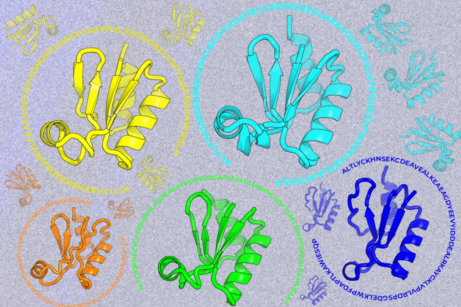
A new machine-learning framework aims to improve the success rate of computational protein design while moving away from results that reproduce sequences found in nature.