Aug 2026· Journal of Advanced Research· 0 citations· 42 references
Medicine
TL;DR
Endothelial DANCR deficiency promotes atherosclerotic plaque instability through activation of the RPL22/p53 pathway, suggesting DANCR as a potential protective factor and therapeutic target in atherosclerosis.
Abstract
INTRODUCTION
Long noncoding RNAs (lncRNAs) are key regulators of vascular endothelial function. The lncRNA differentiation antagonizing non-protein coding RNA (DANCR) is implicated in cell proliferation and inflammatory responses; however, its specific role in atherosclerosis remains undefined.
Objective
To investigate the role of DANCR in modulating endothelial adhesive capacity and atherosclerotic plaque instability.
Methods
DANCR expression was profiled in vascular tissues and cell lines using RNA fluorescence in situ hybridization and real-time qPCR. Endothelial-specific DANCR-knockout mice were generated and injected with recombinant adeno-associated virus carrying murine PCSK9 to induce atherosclerosis. Chromatin isolation by RNA purification followed by sequencing was performed to identify potential targets of DANCR. Plasma DANCR level was measured in healthy subjects (n = 42) and in patients with mixed plaques detected by coronary computed tomography angiography (n = 30).
Results
DANCR expression was predominantly expressed in endothelial cells and was significantly lower by 43% in the endothelium of human carotid plaques than normal vessels. Endothelial-specific knockout of DANCR in mice led to a 78% increase in aortic plaque area and a 1.1-fold elevation in the plaque instability index, characterized by enlarged necrotic cores, elevated type III/I collagen ratio, and increased macrophage infiltration. The ribosomal protein L22 (RPL22) was identified to be a target of DANCR which repressed its transcriptional expression. Endothelial-specific knockdown of RPL22 reversed the plaque progression and instability induced by DANCR deficiency in vivo. In vitro, DANCR reduced endothelial adhesion capacity and the expression of ICAM1 and VCAM1 via inhibition of the RPL22/p53 pathway. Clinically, plasma DANCR level was lower in patients with mixed plaques compared with control subjects.
Conclusions
Endothelial DANCR deficiency promotes atherosclerotic plaque instability through activation of the RPL22/p53 pathway, suggesting DANCR as a potential protective factor and therapeutic target in atherosclerosis.
BACKGROUND
The integrated regulation of microRNAs on macrophage plasticity plays a key role in atherosclerosis. We tested the hypothesis that miR487a-3p and miR6855-3p accelerate atherosclerosis by intensifying macrophage inflammatory response and metabolic dysregulation.
METHODS
The microRNA sequencing and messenger RNA sequencing were conducted in peripheral monocytes from patients with coronary artery disease (CAD) and healthy controls. Macrophages in mouse aortas and human coronary arteries were characterized using flow cytometry and immunostaining. Atherosclerosis development was evaluated in male PCSK9 (proprotein convertase subtilisin/kexin type 9)-overexpression mice harboring myeloid cell-specific deficiency of CPE (carboxypeptidase E) or RRM2 (ribonucleotide reductase regulatory subunit M2) and challenged with a high-fat diet.
RESULTS
miR487a-3p and miR6855-3p were the top microRNA candidates identified by microRNA sequencing in peripheral monocytes and validated by quantitative real-time polymerase chain reaction, with significant differences between patients with CAD and controls. Both microRNAs were lipid-inducible and secreted extracellularly. KLF (Krüppel-like factor 5) and IRF1 (interferon regulatory factor 1) bound to the promoter regions of miR487a-3p and miR6855-3p, respectively, to enhance their transcription. Accordingly, patients with CAD exhibited significantly elevated plasma miR487a-3p and miR6855-3p levels compared with controls, which positively correlated with blood lipid levels and Gensini score (reflecting CAD severity and prognosis). The area under the receiver operating characteristic curve (≈0.83 for each) supported their diagnostic accuracy. Of note, miR487a-3p and miR6855-3p were predominantly expressed in coronary arterial macrophages. The dramatic expansion of miR487a-3p+ and miR6855-3p+ macrophages and the elevated expression of both microRNAs in coronary arteries were positively associated with lesion area in patients with CAD. Mechanistically, transcriptomic analyses and functional assays revealed that elevated miR487a-3p or miR6855-3p promoted macrophage proinflammatory responses, lipid metabolic dysregulation, and foam cell formation. Conversely, inhibition of either microRNA alleviated ox-LDL (oxidized low-density lipoprotein)-induced macrophage inflammatory responses and lipid metabolic dysfunction. Moreover, conditioned medium from miR487a-3p- or miR6855-3p-overexpressing macrophages promoted endothelial cell apoptosis, whereas this effect was attenuated when endothelial cells were exposed to medium from ox-LDL-treated macrophages with microRNA inhibition. Furthermore, integration of downregulated genes from monocyte and macrophage messenger RNA sequencing with TargetScan-predicted targets identified CPE and RRM2 as targets of miR487a-3p and miR6855-3p, respectively. Direct binding was confirmed by dual-luciferase assays and microRNA pulldown. Overexpression of CPE or RRM2 partially reversed the detrimental effects of miR487a-3p and miR6855-3p, respectively, on macrophage phenotypic switching and metabolic dysregulation. Conversely, monocyte-/macrophage-specific depletion of CPE or RRM2 aggravated atherosclerosis progression in hypercholesterolemic mice by instigating macrophage inflammatory responses and lipid metabolic disturbance.
CONCLUSIONS
miR487a-3p and miR6855-3p fulfill the criteria of promising biomarkers for CAD diagnosis and prognosis. Mechanistically, they intensify inflammatory responses and disrupt lipid metabolism in macrophages, identifying both microRNAs as potential therapeutic targets for CAD.
Haijing Ge, Duo Xu, Tao He et al.· Arteriosclerosis, Thrombosis...· 0 citations
Although approximately 97% of the human genome does not encode proteins, it produces a vast repertoire of regulatory RNA transcripts that play critical roles in cellular and molecular processes. Among these, noncoding RNAs including microRNAs and long noncoding RNAs have emerged as key regulators of gene expression through their interactions with DNA, RNA, and proteins. Increasing evidence indicates that noncoding RNAs are central to immune regulation and inflammatory signaling, positioning them as important contributors to complex chronic diseases. Chronic kidney disease (CKD) is of particular interest because it represents a growing global health burden with strong inflammatory and metabolic overlap with cardiovascular disease, placing it at the center of cardio-kidney-metabolic disease. In both atherosclerosis and CKD, a pro-inflammatory milieu characterized by sustained macrophage accumulation and activation drives vascular dysfunction, organ ischemia, and progressive tissue injury. Macrophages act as pivotal instigators of these cardio-kidney-metabolic pathologies by integrating inflammatory signaling, lipid handling, and tissue remodeling processes. This review discusses current knowledge on the roles of noncoding RNAs in regulating macrophage function in atherosclerosis and CKD, with particular emphasis on the molecular mechanisms underlying inflammatory responses and disease progression. By highlighting shared and disease-specific noncoding RNA–mediated pathways, this review aims to provide insight into novel regulatory networks and potential therapeutic targets in cardio-kidney-metabolic disease.
P. Jha, Caio Borges Nascimento, Adrien Lupieri et al.· Frontiers in Cardiovascular...· 0 citations
Mechanistically, circPSEN1 acts as a sponge for miR-150-5p, enhancing TRIM65 expression and contributing to DR pathogenesis, indicating that circPSEN1 may serve as a therapeutic target for DR.
BACKGROUND
Atherosclerotic cardiovascular disease (ASCVD) has shared genetic susceptibility across various vascular lesions. Identification of common pathogenic factors for multi-territorial atherosclerosis is urgently required. We hypothesize that PR domain zinc finger protein 5 (PRDM5) acts as a shared pathogenic regulator to facilitate systemic atherosclerotic lesions by modulating monocyte-macrophage function.
METHODS
Multi-trait genome-wide association studies (GWAS) datasets were analyzed via genomic structural equation modeling (Genomic SEM) to uncover shared risk loci underlying ASCVD. Transcriptome-wide association study (TWAS) was performed in CD14+ monocytes to prioritize candidate genes. PRDM5 expression was validated in clinical peripheral blood monocytes and atherosclerotic plaques. Cellular functional assays, chromatin Immunoprecipitation quantitative real-time PCR (ChIP-qPCR), dual-luciferase reporter assay and molecular docking were combined to decipher downstream signaling and ezetimibe-mediated therapeutic effects.
RESULTS
PRDM5 was identified as a pivotal ASCVD risk gene, with significantly upregulated expression in monocytes and plaque tissues from patients. Mechanistically, PRDM5 directly binds the ASK1 promoter to trigger ASK1/JNK/NF-κB signaling, aggravating macrophage lipid overloading, inflammatory activation and apoptosis, and further inducing endothelial dysfunction through paracrine mediators. Ezetimibe suppresses the transcriptional activity of PRDM5.
CONCLUSION
PRDM5 serves as a common susceptibility gene for ASCVD and facilitates macrophage pathogenic phenotypes via the ASK1/JNK/NF-κB cascade.
Meng Sun, Han Sun, Hanzheng Wang et al.· Experimental Cell Research· 0 citations
ABSTRACT Aortic dissection (AD) is a life‐threatening vascular disorder characterized by separation of the layers of the aortic wall, primarily triggered by an intimal tear or an intramural hemorrhage. Age is a well‐recognized and critical risk factor for AD. Epidemiological evidence consistently demonstrates a strong positive correlation between age and AD incidence, with peak occurrence in the 50–70‐year age group. Its pathogenesis involves vascular inflammation, extracellular matrix (ECM) degradation, vascular smooth muscle cell (VSMC) phenotypic switching, apoptosis, and immune cell infiltration. Two major pathophysiological theories have been proposed: one suggests a primary intimal tear, and the other involves medial degeneration from stress concentration. Long noncoding RNAs (lncRNAs) are transcripts longer than 200 nucleotides and account for ~80% of the human noncoding transcriptome. They exert diverse biological functions (e.g., miRNA sponging, protein scaffolding, transcriptional regulation) and are pivotal in diseases such as cancer, rheumatoid arthritis, and AD. Despite emerging evidence linking lncRNAs to AD, most studies to date have focused only on their role as competing endogenous RNAs (ceRNAs), with limited mechanistic depth and scarce clinical validation. This review summarizes the pathophysiology of AD and systematically discusses lncRNA‐mediated mechanisms in AD progression. We highlight current knowledge gaps and propose future directions for exploring lncRNAs as potential therapeutic targets to improve AD prognosis.
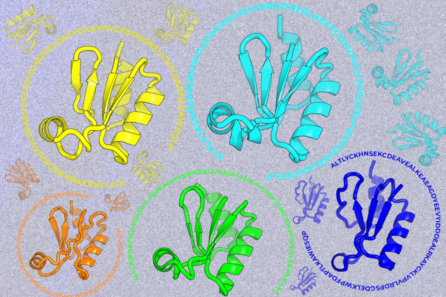
A new machine-learning framework aims to improve the success rate of computational protein design while moving away from results that reproduce sequences found in nature.