This is the first study that provides direct in vitro evidence of Don induced cell cycle arrest at G2/M phase through highly upregulated p21, with no increase in p53 protein levels, suggesting p53-independent p21 induction.
Abstract
Humans are constantly exposed to mycotoxins primarily through contaminated food. Deoxynivalenol (DON) is the most frequently occurring, and its toxicity to stem cells remains unclear. Mesenchymal stem cells (MSCs), responsible for tissue regeneration, serve as tools for regenerative therapies and valuable models for studying cellular differentiation and toxicological responses. This study investigated the cytotoxic effects of DON on the cell cycle progression and differentiation in human dental pulp stem cells (DPSCs). DPSCs, isolated from healthy embedded third-molar teeth and characterized, were treated with DON (0.25-16 µg/mL). Cell viabilities were determined via MTT assay and the inhibitory concentrations (IC50 and IC25) of DON were calculated (0.46 μg/mL and 0.23 μg/mL). Cell death, ROS levels, cell cycle progression, and adipogenic and osteogenic differentiation of DPSCs were evaluated under DON exposure. DON did not induced cell death or oxidative stress in DPSCs. However, DON induced cell cycle arrest at G2/M phase through highly upregulated (6-fold) p21, with no increase in p53 protein levels, suggesting p53-independent p21 induction. Moreover, DON increased the expression of early differentiation markers (BMP2 and C/EBPβ) under non-induced culture conditions. This is the first study that provides direct in vitro evidence of DON on the cell cycle progression and differentiation in DPSCs.
Investigating the cytotoxic effects of DON and its underlying mechanisms using a 3D spheroid model derived from the human neuroblastoma cell line SH-SY5Y demonstrates that DON induces cytotoxic effects in SH-SY5Y spheroids, accompanied by mitochondrial alterations, increased mitochondrial superoxide production, and apoptotic cell death.
M. Sánchez, Zingales, J. Tolosa et al.· Toxicology· 0 citations
Introduction: Diabetes mellitus (DM) being a chronic metabolic disorder, causes a major concern for the healthcare system. Among different types, type 1 DM (T1DM) results in the destruction of insulin-producing pancreatic β cells, mediated by the immune system. Studies have demonstrated that human umbilical cord derived mesenchymal stem cells (hUMSCs) exhibit great potential to regenerate β-cells. Moreover, in order to enhance the regenerative potential of MSCs, several strategies are being utilized, including preconditioning with bioactive compounds. Among these, naphthoquinones can be used for MSC preconditioning in order to augment their therapeutic potential for β-cell regeneration, as these compounds possess anti‐inflammatory and anti-diabetic properties. Methods: hUMSCs were isolated, characterized, and treated with non-cytotoxic concentrations of lawsone, lapachol, or their combination. The preconditioned cells were subsequently analyzed for pancreatic β-cell differentiation at gene and protein levels. The study also explores the role of Wnt and BMP signaling pathways during the differentiation process through gene expression analysis. Binding patterns of these compounds with their respective receptors were analyzed using in silico studies. Results: Gene expression profiling showed overexpression of pancreatic β-cell–specific markers in the Law + hUMSC group, whereas downregulation of Neurogenin-3 (NGN3) was observed in all treatment groups. Immunocytochemical analysis also showed enhanced expression of insulin in Law + hUMSCs, relative to other groups. Transcriptional analysis of the wingless/integrated (Wnt) and bone morphogenetic protein (BMP) pathways showed increased Wnt and decreased BMP expression across all treatment groups. In silico analyses showed that the binding patterns of lawsone or lapachol with frizzled (FZD) and activin-like kinase 1 (ALK1) receptors share comparable sequence similarity, facilitating their binding to these receptors and regulating downstream Wnt and BMP signaling. Conclusion: The study concludes that the regulatory role of lawsone and lapachol can be exploited for preconditioning of MSCs for improved pancreatic β-cell differentiation.
HHT and Tan IIA combined therapy synergistically suppressed lymphoma progression by downregulating the Akt/MMP signaling pathway, suggesting their use in treating lymphomas.
Yanping Zhang, Jian Yin, Rui Liu et al.· Cytotechnology (Dordrecht)· 0 citations
ADSC-derived CINVs effectively regulate the biological functions of DPCs and promote the transition of hair follicles from telogen to anagen, primarily through activation of the β-catenin signaling pathway.
Lei Wang, Yiwen Liu, Peiyun Cai et al.· Zhejiang da xue xue bao. Yi...· 0 citations
DA elicits a biphasic cellular response in U937 leukemia cells, characterized by transient cytoprotective autophagy followed by apoptotic cell death mediated through dysregulation of Bcl-2/Bax and suppression of the PI3K/Akt pathway, indicating that DA is a promising candidate for novel antileukemic therapeutic strategies.
Cancer progression involves intricate interactions between inflammatory signaling, programmed cell death mechanisms, and oxidative stress. Although enoxaparin is widely used for managing cancer-associated thrombosis, its direct cellular effects on tumor biology remain insufficiently characterized. This study aimed to evaluate the impact of enoxaparin on apoptosis, autophagy, inflammatory mediators, and oxidative DNA damage in breast (MDA-MB-231) and liver (HepG2) cancer cell lines. MDA-MB-231, HepG2, and non-cancerous HEK-293 cells were treated with varying concentrations (5, 10, 20, 40, and 80 mg/mL) of enoxaparin for 24 and 48 h. Cell viability was assessed using the MTT assay, while apoptosis was quantified by TUNEL analysis. Immunofluorescence staining was employed to evaluate the expression of NF-κB, IL-6, TNF-α, LC3, and p62. Oxidative DNA damage was determined by measuring extracellular 8-hydroxy-2'-deoxyguanosine (8-OHdG) levels using a competitive ELISA. Statistical analyses were conducted to compare the treated and control groups. Enoxaparin significantly reduced cell viability in MDA-MB-231 and HepG2 cells without inducing cytotoxicity in HEK-293 cells. Apoptosis was markedly increased in both cancer cell lines following treatment. Enoxaparin differentially modulated inflammatory signaling; NF-κB expression was significantly increased in MDA-MB-231 cells, accompanied by suppression of IL-6 and TNF-α, whereas no significant inflammatory changes were observed in HepG2 cells. Enoxaparin treatment was observed to increase LC3 and p62 expression in both MDA-MB-231 and HepG2 cells, triggering autophagy-related pathways. Moreover, enoxaparin significantly reduced extracellular 8-OHdG levels, suggesting a reduction in oxidative DNA damage. Enoxaparin exhibits multifaceted anticancer effects by promoting apoptosis and autophagy, selectively modulating inflammatory pathways, and reducing oxidative DNA damage in breast and liver cancer cells.
Sedat Çarkıt, M. Baran, N. Bitgen et al.· Scientific Reports· 0 citations
Related blog posts
MIT News · Artificial Intelligence· news.mit.eduAug 27, 2026
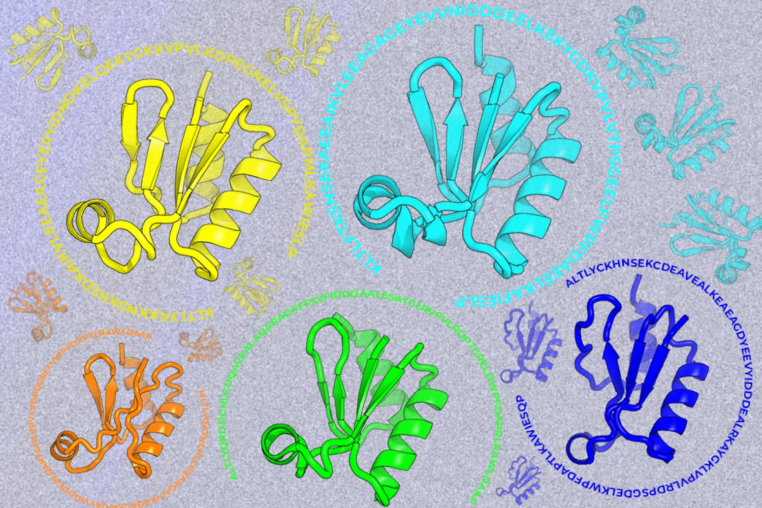
A new machine-learning framework aims to improve the success rate of computational protein design while moving away from results that reproduce sequences found in nature.