Aug 2026· Iranian Journal of Pathology· Vol 21, pp. 560 - 570· 0 citations· 31 references
Medicine
TL;DR
A divergent regulation of CD29 and CD49b integrins associated with HLA-B27 status in Iraqi AS patients is suggested and longitudinal validation is required to confirm their clinical utility as diagnostic or therapeutic targets.

Abstract
Background & Objective: Ankylosing spondylitis (AS) is a systemic inflammatory disorder where the HLA-B27 allele significantly influences disease severity. This study evaluated the molecular profiles of integrins CD29 and CD49b in peripheral blood mononuclear cells (PBMCs) to identify potential biomarkers associated with HLA-B27 status. Methods: In this cross-sectional study of an Iraqi cohort, 130 participants were recruited: 50 HLA-B27-positive (HLA+) radiographic AS patients, 50 HLA-B27-negative (HLA−) radiographic AS patients, and 30 healthy controls. Transcriptomic and proteomic expressions of CD29 and CD49b were quantified using qPCR and flow cytometry (mean fluorescence intensity, MFI). Hematological and biochemical profiles were also assessed. Results: CD29 mRNA was significantly upregulated in both patient groups, with a more pronounced median elevation in the HLA+ group (11-fold; P < 0.0001) than in the HLA− group (3-fold; P < 0.001). Surface CD29 protein significantly increased only in the HLA+ cohort (mean MFI approximately 3000; P < 0.0001). Conversely, CD49b mRNA was substantially suppressed, particularly in the HLA+ group (P < 0.0001). Correspondingly, a marked reduction in CD49b surface protein occurred in the HLA+ cohort (mean MFI approximately 300; P < 0.0001), whereas the HLA− group remained statistically comparable to controls. Additionally, the HLA− phenotype was characterized by significant granulocytosis (85%–90%) and more pronounced elevations in urea and ALT levels. Conclusion: These findings suggest a divergent regulation of CD29 and CD49b integrins associated with HLA-B27 status in Iraqi AS patients. While these integrins may serve as molecular indicators for disease stratification, longitudinal validation is required to confirm their clinical utility as diagnostic or therapeutic targets.
Ankylosing spondylitis (AS) is a chronic, immune-mediated inflammatory disorder primarily affecting the axial skeleton. It is characterized by bony fusion and syndesmophyte formation of the spine, leading to compromised mobility, reduced quality of life and increased mortality. However, the underlying immune dysregulation in AS remains poorly understood. We comprehensively profiled the peripheral immune landscape of 30 patients with active AS, all of whom were TNF-α blocker-naïve, capturing the immune profile during the active disease phase without biologic interference. Using advanced 11-color flow cytometry and FlowSOM clustering, 584 immunophenotypes were analyzed in AS and 30 age-and sex-matched healthy controls, with emphasis on both cellular composition and functional characteristics. AS patients displayed a distinctive functional “activated-exhausted” immune phenotype, dominated by CD4
+
CD28
+
CD38
+
PD-1
+
T cells and HLA-DR
+
CD38
+
CD4
−
CD8
−
double-negative (DN) T cells. DN T cells were significantly enriched in HLA-B27-positive patients, suggesting potential as a biomarker. Network analysis revealed seven central immune subsets associated with active disease, while functional assessment demonstrated impaired CD95
+
CD8
+
T cell apoptosis and disrupted B-cell maturation. These findings provide a detailed functional immune map of biologic-naïve active AS, offering novel mechanistic insights and identifying candidate biomarkers for precision intervention.
Lu Bai, Jian Gao, Yunyun Xu et al.· Phenomics· 0 citations
Behçet's disease is an uncommon multisystem inflammatory condition characterized by immune-mediated vasculitis affecting vessels of different calibers. Clinically, it presents with recurrent oral and genital ulcerations, dermatological manifestations, and ocular involvement, reflecting its systemic nature. The present study was designed to characterize gut microbial patterns in patients diagnosed with Behçet’s disease and to assess selected immune biomarkers — IL-17A, IL-23, CD4, and CD11b — as potential diagnostic and prognostic indicators. Participants were recruited from Al-Sadr Medical City between December 2024 and September 2025, with ages ranging from 11 to 51 years. Venous blood samples were obtained from patients, and serum concentrations of IL-17A, IL-23, CD4, and CD11b were quantified using the ELISA technique. The study population showed a predominance of male patients, with the highest frequency observed in the 22–31-year age group. Statistical analysis demonstrated significantly elevated serum levels of IL-17A and IL-23 in Behçet’s patients compared with healthy controls. Similarly, CD4 and CD11b expression levels were markedly increased. A strong positive correlation was identified among these immune markers, suggesting coordinated immunological activation. In conclusion, IL-17A, IL-23, CD4, and CD11b may serve as valuable diagnostic and predictive biomarkers in Behçet’s disease, and their interrelationship highlights their potential role in disease pathogenesis and progression.
T cells play a central role in axial spondyloarthritis (axSpA). The serine-threonine kinase DRAK2, encoded by STK17B, modulates T cell activation and survival. In autoimmune models, it promotes autoreactive T cell persistence and reduces regulatory T cell (Treg) abundance.[1,2] We previously found STK17B upregulation in peripheral blood mononuclear cells (PBMCs) of axSpA patients who were non-responders to tumor necrosis factor inhibitors (TNFi).[3] To clarify whether this reflects disease-associated inflammation or treatment resistance, we compared STK17B/DRAK2 expression (i) between axSpA patients and healthy controls and (ii) between responders and non-responders to IL-17 inhibitor (IL-17i) therapy. We also examined STK17B/DRAK2 expression in the SKG mouse model of SpA.
Human spinal tissue from axSpA patients and spinal trauma controls was analyzed by immunohistochemistry (IHC) to localize DRAK2 expression, and by immunofluorescence (IF) to compare expression between CD4
+
and CD8
+
T cells. PBMCs from patients and controls were sorted by fluorescence-activated cell sorting (FACS), and STK17B expression in each subset was quantified by qPCR. Parallel IHC was performed on ankle, tail, and ileum of curdlan-treated and PBS-treated SKG mice. To corroborate findings at higher resolution, STK17B expression in mature CD4
+
and CD8
+
T cells was queried from our pre-biologic single-cell RNA-sequencing and multiome datasets.
In human spinal tissue, DRAK2
+
immune infiltrates were more abundant in axSpA than in controls (Figure 1A), with stronger IF staining in CD4
+
than CD8
+
T cells. In PBMCs, STK17B was preferentially expressed in CD4
+
T cells and trended higher in axSpA compared with controls. Single-cell and multiomic analyses confirmed this pattern, showing modest STK17B increases in CD8+ T cells but markedly higher levels in CD4+ T cells of axSpA patients, particularly in effector T cells and Tregs. Notably, pre-biologic STK17B expression was higher among patients who were later identified as non-responders to IL-17i compared with responders (Figure 1B). Consistent with the human findings, curdlan-treated SKG mice exhibited increased DRAK2
+
infiltrates in ankle, tail, and ileum relative to PBS-treated controls, paralleling the observation in human tissues (Figure 1C).
Figure 1.
(A) Histopathology of spinal tissues from AS and control patients with IHC staining for anti-DRAK2. (B)
STK17B
expression in mature CD4+ T cells of pre-biologic axSpA patients and controls. (C) Histopathology of the tissues from curdlan- vs PBS-SKG mice for anti-DRAK2.
Across humans and mouse models, STK17B/DRAK2 expression is increased in inflammatory lesions and enriched in CD4
+
effector and regulatory T cells, particularly in patients with inadequate IL-17i response. These findings suggest that DRAK2 upregulation contributes both to axSpA-related inflammation and to treatment resistance, warranting further mechanistic investigation.
[1.] Ramos S. J Immunol 2008;181:7606-16. [2.] Mandarano A. Cell Rep 2023;42:112106. [3.] Talukdar A. [Abstract]. Arthritis Rheumatol 2024;76 Suppl 9.
P. Remalante-Rayco, S. Jo, Addison Pacheco et al.· Journal of Rheumatology· 0 citations
Peripheral GZMB+CD4+ CTLs are expanded in DM and ASS and are closely associated with disease activity, while their diagnostic performance suggests that they may serve as a biomarker and potential therapeutic target in these diseases.
Qin Zhang, Rui Chi, GuoXun Shi et al.· Frontiers in Immunology· 0 citations
Serum sCD40 does not reflect disease activity or the humoral immune response in rheumatoid arthritis and further studies are needed to evaluate its relevance during early stages of the disease and to further characterize its role in cytokine production.
Valeria Miroslava Limón-López, J. Muñóz-Valle, Zyanya Reyes-Castillo et al.· Journal of Clinical Medicine· 0 citations
Related blog posts
MIT News · Artificial Intelligence· news.mit.eduAug 27, 2026
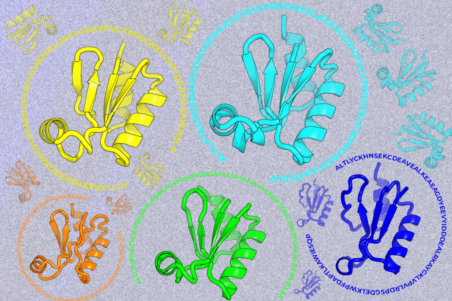
A new machine-learning framework aims to improve the success rate of computational protein design while moving away from results that reproduce sequences found in nature.