A Site-Specific
Organometallic Approach for Installing
Tyrosine Phosphorylation Mimics to Decipher the Role of Phosphorylation
in α-Synuclein Aggregation and Seeding
Aug 2026· Journal of the American Chemical Society· 0 citations· 65 references
TL;DR
This strategy provides a rapid, selective, and scalable way to introduce aromatic PTMs, producing homogeneous protein libraries essential for mechanistic studies, biomarker development, and exploring the therapeutic potential of targeting aSyn-phosphorylation for PD and related disorders.

Abstract
Several studies have identified phosphorylation of α-synuclein (aSyn) at multiple tyrosine residues (Y39, Y125, Y133, and Y136) within Lewy bodies (LB), which are the pathological hallmarks of Parkinson’s disease (PD). However, understanding the specific role of phosphorylation at each site, or how multiple phosphorylation sites interact, has been challenging. Herein, we present an efficient method that leverages an organometallic Pd-complex to site-specifically attach phosphomimetic groups via Cys-arylation, closely mimicking natural phosphorylation. Using this approach, we successfully incorporated native-like tyrosine phosphorylation mimics into cysteine-containing proteins like synthetic transcription factor Max and recombinant aSyn, in good yields. To demonstrate the method’s versatility, we created a focused library of mono-, di-, and triphosphorylated aSyn analogues. This development enabled, for the first time, the investigation of the effect of site-specific phosphorylation at multiple C-terminal tyrosine residues on aSyn fibrillization and aggregation. Our results show that monophosphorylation at any of the C-terminal sites has little effect on aggregation, while di- and triphosphorylation slows down the process, extending the lag phase compared to wild-type aSyn. Additionally, phosphorylation at Y39 reduces the seeding activity of aSyn fibrils by almost 2–4-fold in both mammalian and neuronal models of synuclein pathology. Overall, our strategy provides a rapid, selective, and scalable way to introduce aromatic PTMs, producing homogeneous protein libraries essential for mechanistic studies, biomarker development, and exploring the therapeutic potential of targeting aSyn-phosphorylation for PD and related disorders.
Intrinsically disordered proteins (IDPs) drive many neurodegenerative disorders, but their structures remain difficult to define because they populate dynamic ensembles that change with the chemical environment. This problem is central for α-synuclein (aSyn), a Parkinson’s disease-linked IDP in which S129 phosphorylation is highly enriched in disease-associated aggregates. Although S129 phosphorylation stabilizes a more compact aSyn ensemble in dilute solution, whether this structural effect persists across other biochemical environments has remained unknown. Here, we developed a quantitative cross-linking mass spectrometry framework to determine how WT and pS129 aSyn respond to chemically distinct environments. We compared dilute buffer with two perturbations relevant to aSyn biology: trimethylamine N-oxide (TMAO), a gut-microbiome-derived metabolite associated with Parkinson’s disease that can also act as a compacting osmolyte at high concentration, and octyl glucoside (OG) micelles, which provide a membrane-mimetic surface. TMAO rewired the phosphorylation-dependent structural response in a concentration-dependent manner: 1.8 M TMAO shifted WT aSyn toward the dilute pS129 contact pattern by increasing long-range contacts between the N-terminal and C-terminal regions, whereas 3.4 M TMAO collapsed both proteoforms and reduced their structural differences. In OG micelles, the proteoforms diverged. WT favored an extended-helix-like contact pattern with stronger contacts between the C-terminal tail and micelle-bound N-terminal region, whereas pS129 favored contacts between the N-terminal and NAC regions and fewer contacts to the C-terminal tail, consistent with a broken-helix-like topology. By integrating regional contact counts, normalized cross-link intensities, and geometric compatibility analysis, this workflow distinguishes broad contact accessibility from the residue-level contacts that dominate each condition. These results establish a proteoform-by-environment model for IDP structure, in which phosphorylation effects are not fixed but are rewritten by the surrounding chemical environment.
Ashlyn N. Dollar, Daniel W. Kelley, Ian K. Webb· Journal of the American Chem...· 0 citations
Protein tyrosine phosphorylation is critical for cellular function, and aberrant phosphorylation is tied to a wide range of human diseases. Identifying the substrates of protein tyrosine phosphatases, the enzymes that erase this modification, is critical to understanding human biology and disease states. The state-of-the-art method for tyrosine phosphatase substrate identification requires the use of mutations that modestly increase the lifetime of enzyme-substrate complexes by kill catalytic activity. While these “substrate-trapping” mutants are useful tools, they work best for high-affinity or abundant substrates that remain phosphatase-bound through cell lysis and enrichment. Here, we use site-specific photo-crosslinking to covalently capture the substrates of tyrosine phosphatases in situ. We identify eight different positions around the active site of the phosphatase PTP1B where photo-crosslinker amino acids can be incorporated via amber codon suppression without dramatically disrupting catalytic activity. We then conduct photo-crosslinking experiments in mammalian cells and identify crosslinked proteins by mass spectrometry proteomics, revealing that our approach can capture known PTP1B interactors and substrates. We then show that PTP1B photo-crosslinking in situ is sensitive to enzyme localization and identify new PTP1B substrates that regulate contacts between the endoplasmic reticulum and plasma membrane. We also demonstrate that photo-crosslinking can capture signal-dependent interactions. For example, we observe PTP1B crosslinking to the epidermal growth factor (EGF) receptor, a known substrate, in an EGF-dependent manner, and we identify other potential EGF-dependent substrates. Overall, our approach reveals previously unknown roles of PTP1B in signaling systems and could be readily extended to other tyrosine phosphatases in the same family.
Andrew C. Johns, Yethmie S. Goonatilleke, David C. Cabanero et al.· bioRxiv· 0 citations
α‐Synuclein (αSyn) is a major component of pathogenic Lewy bodies and Lewy neurites and is closely associated with Parkinson's disease. Among the various posttranslational modifications of αSyn, several have been implicated in the degeneration of dopaminergic neurons and are thought to promote Parkinson's disease through enhanced misfolding, aggregation, and accumulation of αSyn. Two such modifications, phosphorylation at Ser129 (S129Phos) and hydroxylation at Tyr136 (Y136DOPA), exert distinct effects on αSyn aggregation: S129Phos has been reported to either inhibit or promote aggregation, whereas Y136DOPA induces the formation of short oligomeric species. To gain insight into the molecular basis underlying the initiation of αSyn multimerization, we semisynthesized αSyn carrying either S129Phos or Y136DOPA and prepared recombinant unmodified full‐length αSyn as a control. Vacuum‐ultraviolet circular dichroism (VUVCD) spectroscopy revealed that these αSyn variants in their monomeric states possessed essentially identical secondary structures. These results suggest that the modifications themselves do not induce significant secondary structural changes in monomeric αSyn.
Tatsuhito Matsuo, I. Suetake, Mariko Kimura et al.· Journal of Peptide Science· 0 citations
Site-specific phosphorylation controls protein function, interactions, and cellular fate, but the effects of individual phospho-proteoforms remain difficult to define in cells. Existing methods rely on kinase-mediated phosphorylation, which often generates heterogeneous mixtures that are phosphatase-sensitive, or on phosphomimetic substitutions that frequently fail to reproduce authentic phospho-states. Here we establish direct phospho-proteoform delivery, which combines genetic code expansion-enabled production of proteins containing the nonhydrolyzable phosphoserine analog nhpS in E. coli with electroporation-based delivery of purified proteins into human cells. This strategy creates an experimental framework for testing defined phospho-proteoforms in cells with control over identity, dose, and timing, independent of intracellular kinase, phosphatase, and expression systems. Using this capability, we tested whether phosphorylation-induced monomerization of 14-3-3 proteins is sufficient to alter cellular protein fate, a question that cannot be addressed by conventional methods. Installation of nhpS at the conserved dimer interface created a proteasome-sensitive, degradation-prone state across multiple paralogs. Although cereblon preferentially associated with monomeric 14-3-3, cereblon knockout did not rescue degradation, and monomeric 14-3-3 also engaged the E3 ligase adaptor SKP1. These findings reveal phosphorylation-induced monomerization as a conserved trigger of 14-3-3 proteostatic control and establish direct phospho-proteoform delivery as a general route to connect single phosphorylation events to cellular phenotypes.
Moriah H. Mathis, Stanislau Stanisheuski, Ryan A. Mehl et al.· Chemical Science· 0 citations
Catalytic activity of 5′-3′ exonuclease Phospholipase D3 (PLD3) is associated with immune signaling and neurodegeneration including Alzheimer’s disease. PLD3 undergoes multiple post-translational modifications and proteolytic cleavage to establish its catalytically active form. However, the proteases catalyzing the cleavage of PLD3 have remained unidentified. To study the proteolytic cleavage of PLD3, we have evaluated the small molecule covalent inhibitor E64d that blocks proteolysis catalyzed by cysteine cathepsins. To validate the selectivity of E64d, we have designed and synthetized an E64d propargyl analogue and carried out a detailed activity-based protein profiling to reveal a broad engagement of the compound with other protein targets including bleomycin hydrolase (BLMH), Kelch-like ECH-associated protein 1 (KEAP1), transcription elongation factor SPT5 (SUPT5H) and asparagine synthetase (ASNS). The specificity of the E64d-protein interactions was confirmed by biochemical assays and mass spectrometry-based site identifications. In neurons, treatment with E64d lead to about 50-fold PLD3 accumulation and dysregulation of its proteolytic cleavage, while there was only a minor overall change on the whole proteome level. Taken together, this study provides insights into previously unknown E64d selectivity and renders cysteine cathepsins responsible for PLD3 degradation in neurons. It highlights the importance of cysteine cathepsins activity in neuronal lysosomes for proper PLD3 processing and hence it suggests that their activation might be responsible for decreased PLD3 levels in neurons of patients with Alzheimer’s diseases. These findings are key for further elucidation of PLD3 function in neurodegenerative diseases.
Manuel Hertwig, P. Kielkowski· bioRxiv· 0 citations
Related blog posts
MIT News · Artificial Intelligence· news.mit.eduAug 27, 2026
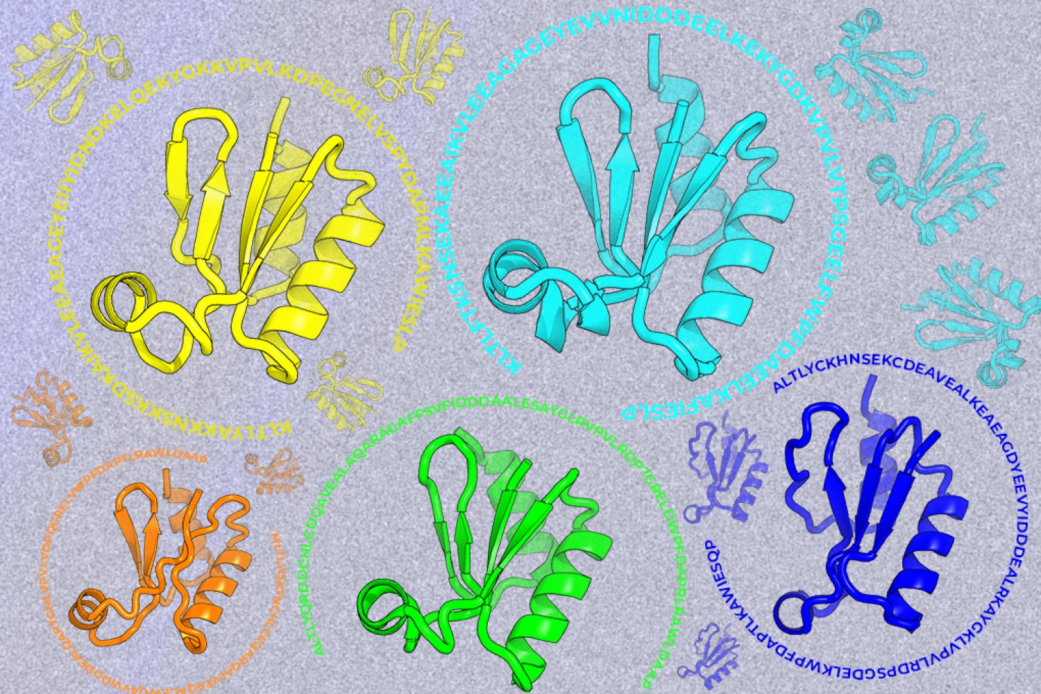
A new machine-learning framework aims to improve the success rate of computational protein design while moving away from results that reproduce sequences found in nature.