Skip to content
#protein folding Open access

The molecular basis of mitochondrial crista formation by the MIC10 complex

Aug 2026 · Science Advances · Vol 12 · 0 citations · 73 references
Medicine

TL;DR

It is found that the MIC10 proteins Mic10, Mic26, and Mic27 strongly recruit cardiolipin at conserved positive loop motifs, driving oligomerization of these subunits and resulting in the stabilization of curvature in model membranes.

Abstract

Mitochondrial cristae are essential for respiration, yet the molecular basis of how the high curvature of these membrane folds is maintained remains unclear. Using structure prediction tools and multiscale simulations, we examined the role of the MIC10 subcomplex of the mitochondrial contact site and cristae organizing system (MICOS). We found that the MIC10 proteins Mic10, Mic26, and Mic27 strongly recruit cardiolipin at conserved positive loop motifs, driving oligomerization of these subunits and resulting in the stabilization of curvature in model membranes. Reconstruction of the full MIC10 complex in a realistic crista junction setup shows its capability to maintain membrane bending, while intrinsically disordered regions may form a permeability barrier between cristae and the intermembrane space. These findings provide a mechanistic model for cristae curvature formation and suggest how MICOS components cooperate with cardiolipins to maintain mitochondrial architecture.

Read PDF

Similar papers

Review Open access Aug 2026

The molecular mechanisms of crista formation: how mitochondria give themselves breathing room.

Cristae are mitochondrial subcompartments that give the organelle its distinctive appearance. More significantly, mitochondria are the proverbial powerhouses as cristae house the molecular machinery underlying cellular respiration, a process that converts carbon sources into ATP by chemiosmosis. The form of cristae is invariably connected to their bioenergetic function. Here, we review our current understanding of the molecules underpinning crista formation. Not surprisingly, respiratory chain multiprotein complexes are involved in crista formation, with F1FO-ATP synthase dimers being eminent membrane sculptors. But crista formation also requires factors that are not directly part of the respiratory chain. The most ancient is the MICOS complex, which delineates the subcompartment and acts as a hub for crista biogenesis. The mitochondrial inner membrane (IM), from which cristae emerge, is remodelled by different dynamin-related proteins in animals and fungi. Cardiolipin is an integral component of the membranous fabric of the IM. To begin to grasp general design principles underlying crista formation, we synthesize findings from canonical animal and yeast experimental models with those from diverse protists and other eukaryotes. However, how these molecules are orchestrated during crista formation remains a hidden piece in our understanding of how cells differentiate in specialized forms. We highlight the few knowns about crista formation in a handful of organisms to guide research into the many unknowns about how complex subcompartments represented by mitochondrial cristae are formed.

Lilia Colina-Tenorio, Martina Bohuslavová, Alexander W. Bruce et al. · 0 citations
Open access Jul 2026

Integrative structure determination of a human mitochondrial contact site and cristae organizing system (MICOS) sub-assembly

The Mitochondrial contact site and Cristae Organizing System (MICOS) complex is an inner mitochondrial membrane (IMM) assembly present at the cristae junction. It is responsible for regulating cristae formation and remodeling. However, its structure is not known. We applied Bayesian integrative structure determination to characterize the structure of the Mic60, Mic19, Mic10, and Mic13-containing MICOS complex combining AlphaFold predictions with data from crosslinking mass spectrometry, biochemical assays, electron tomography, homology modeling, and sequence alignments. The integrative structure revealed novel mutual interfaces among Mic10N,C, Mic60LBS1,LBS2,mitofilin, and Mic13central,C, which were experimentally validated. Several likely-pathogenic missense mutations also localize to these novel interfaces, highlighting their importance. Our results indicate that Mic13 likely facilitates MICOS assembly by binding Mic10 in the IMM-proximal region and Mic60 in the intermembrane space. Taken together, our integrative approach sheds light on the structure and assembly of the MICOS complex.

Muskaan Jindal, Rakesh Mahato, Sreemoyee Das et al. · 0 citations
Open access Jul 2026

Trypanosomal MICOS is assembled on non-respiring mitochondrial crista precursors and associates with two integral microproteins

The mitochondrial contact site and cristae organizing system (MICOS) is a multiprotein complex that shapes crista junctions and maintains inner and outer membrane contacts. MICOS coordinates the assembly of electron transport chain complexes, a prerequisite for cellular respiration. Indeed, MICOS is lost in eukaryotes that dispensed with cellular respiration, suggesting that its assembly depends on the presence of an active respiratory chain. Trypanosoma brucei provides a unique system to test this hypothesis as its mitochondrion undergoes developmentally regulated remodeling. In the insect stage, the mitochondrion contains cristae with an active electron transport chain, whereas the mammalian bloodstream form possesses precursor cristae with stub-like morphology that lack respiratory activity. MICOS has been characterized in the insect stage but remains unexamined in the bloodstream form. Here, we demonstrate that all MICOS subunits assemble onto precursor cristae, retaining conserved interactions with both outer and inner membrane protein machineries. This is somewhat unexpected given the co-occurrence of MICOS with active cellular respiration in nature. Furthermore, we identify novel MICOS-associated proteins that are dispensable for its stability, suggesting auxiliary rather than core roles in MICOS function. Together, our findings establish that MICOS assembly precedes cellular respiratory competence and expand its interaction landscape in trypanosomatids.

Michala Boudová, Teresa Wagner, Tomáš Bílý et al. · 1 citation
Aug 2026

Functional Reconstitution of Mitochondrial Respiratory Supercomplexes.

Mitochondria are central hubs in bioenergetic metabolism and are the primary source of ATP. The inner mitochondrial membrane houses the oxidative phosphorylation system, which includes electron transport chain complexes (CI, CII, CIII2, and CIV) and the ATP synthase (CV). In mammals, CI, CIII2, and CIV form higher-order structures called supercomplexes (SCs) such as SC I+III2+IV, SC I+III2, and SC III2+IV. Although the physiological factors favoring SC formation remain unclear, it has been proposed that SC formation may enhance electron-transfer rates between complexes, reduce reactive oxygen species production, or prevent nonspecific protein aggregation within the densely packed mitochondrial inner membrane. Structural and functional studies of respiratory SCs have relied heavily on detergent-extracted complexes. While these studies have improved our understanding of the electron transport chain, the lack of a sealed membrane bilayer limits their ability to probe the functional benefits of supercomplex assembly. Recent advances, however, have shown that membrane proteins can be structurally characterized in reconstituted, native-like membrane environments, offering a more physiological context for these investigations. Here, we present a simple, quick, and reproducible protocol for reconstituting respiratory SCs into liposomes. This method allows for testing the effects of varying lipid compositions, protein concentration, and membrane potential on the function of respiratory SCs, providing a valuable tool for future mechanistic studies.

A. Padavannil, Peiyi Liu, J. Letts · 0 citations
#protein folding Aug 2026

Structural basis of Saccharomyces cerevisiae Mba1 in mitochondrial co-translational membrane insertion.

The solution structure of mature Saccharomyces cerevisiae Mba1 is determined using multidimensional nuclear magnetic resonance (NMR) spectroscopy and reveals a compact α + β fold with a central hydrophobic cavity and distinct charged surface regions, providing a structural framework for interpreting previous functional studies of Mba1.

Jing Yang, M. Ruan, Dong-Shuai Bai et al. · 0 citations
Review Open access Aug 2026

Mitochondrial Cristae as Separate Compartments: Linking Organization and Function

Traditional bioenergetic paradigms historically relied on classical equilibrium thermodynamics to calculate mitochondrial kinetics, often overlooking the non-equilibrium processes dictated by complex structural architecture. Recent discoveries fundamentally challenge these outdated views by demonstrating that the inner mitochondrial membrane is strictly segregated into distinct functional domains, where individual cristae operate as autonomous, ultra-confined nanocompartments, where the transport of metabolites and protons is tightly controlled by ultrastructure-assisted electric and entropic effects. Compartmentalization prevents proton dissipation, allows for the rapid generation of a localized proton motive force optimized for efficient ATP synthesis and provides robust functional redundancy against localized membrane damage. Furthermore, recognizing cristae as isolated microspaces resolves the long-standing paradox of mitochondrial nicotinamide adenine dinucleotide transhydrogenase (TH). We describe a multi-stage transport pipeline—the TH–isocitrate dehydrogenase axis—wherein matrix-generated reducing equivalents are exported into the cytoplasm via an irreversible isocitrate/α-ketoglutarate loop. This universal pipeline continuously supplies uncommitted NADPH for biosynthesis, systemic antioxidant defense and detoxification. We also highlight the role of compartmentalization in ATP transport and utilization processes. Consequently, disruptions to cristae compartmentalization emerge as primary pathogenic drivers in ischemic, neurodegenerative, and cardiovascular diseases.

A. Panov, S. V. Nesterov, L. Yaguzhinsky · 0 citations

Related blog posts

MIT News · Artificial Intelligence Aug 27, 2026

Looking beyond natural sequences

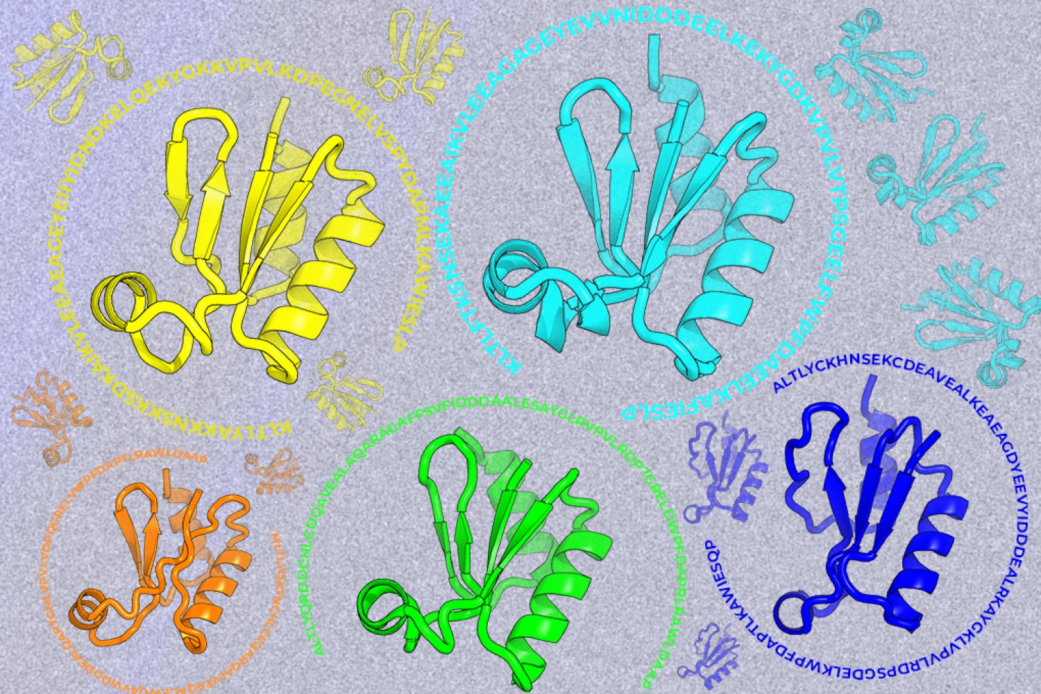
A new machine-learning framework aims to improve the success rate of computational protein design while moving away from results that reproduce sequences found in nature.

Google DeepMind Blog Nov 25, 2025

AlphaFold: Five years of impact

Explore how AlphaFold has accelerated science and fueled a global wave of biological discovery.