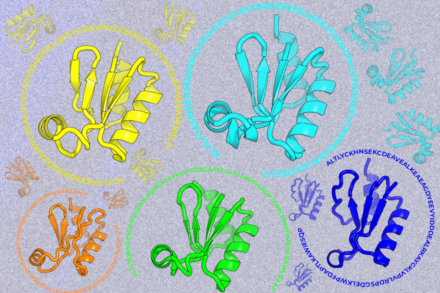
The 1H, 13C, and 15N resonance assignments of the Amblyomin-X Kunitz domain obtained by multidimensional NMR spectroscopy provide the first detailed NMR characterization of the Amblyomin-X Kunitz domain l.
Abstract
Amblyomin-X is a Kunitz-type inhibitor of factor Xa (FXa) from the tick Amblyomma sculptum with promising anticoagulant activity and potential therapeutic applications. Despite the relevance, the molecular basis of its interaction with FXa remains poorly understood, limiting a detailed understanding of its mechanism of action. In this study, we report the 1H, 13C, and 15N resonance assignments of the Amblyomin-X Kunitz domain obtained by multidimensional NMR spectroscopy. High-quality spectra enabled extensive assignment, reaching 97.9% completeness for backbone assignments and 79.8% for side-chain assignments. The 1H-15N HSQC spectrum shows excellent signal dispersion, consistent with a well-folded protein in solution. Furthermore, chemical shift-based structure analysis reveals elements that agree with the canonical Kunitz fold, including characteristic β-strands and helical regions. These results provide the first detailed NMR characterization of the Amblyomin-X Kunitz domain l. The resonance assignments presented here constitute a critical foundation for future structural and dynamical studies, including analyses of protein-ligand interactions. Ultimately, this work contributes to a deeper understanding of the molecular determinants governing FXa inhibition by Amblyomin-X and supports ongoing efforts to develop novel anticoagulant strategies based on Kunitz-type inhibitors.
Protein and nucleic acid alkylation are important genetic and epigenetic modifications. The dynamic balance between alkylation and dealkylation is regulated by distinct sets of enzymes and is essential for maintaining genomic stability. Escherichia coli AlkB is a member of the Alkylation B (AlkB) family of dioxygenases that dealkylates a wide range of nucleic acid substrates in E. coli, thereby playing a crucial role in cellular repair processes and epigenetic regulation. Here, we report the backbone 1H, 15N, 13C chemical shift assignment of E. coli AlkB in complex with Zn2+ and α-ketoglutarate. Experiments were acquired at 20 °C by heteronuclear multidimensional NMR spectroscopy. Collectively, 91% of all 13C, 15N and 1H backbone resonances of Alkb were assigned, with 182 out of a possible 200 residues assigned in the 1H–15N TROSY spectrum. Using the program TALOS + , a secondary structure prediction was generated from the assigned backbone resonances that is consistent with the previously reported X-ray structure of the enzyme. The reported assignment will permit investigations of the protein structural dynamics anticipated to provide crucial insight regarding fundamental aspects in the recognition and enzyme regulation processes.
Sayan Das, J. Purslow, Kyle Malcom et al.· Biomolecular NMR Assignments· 0 citations
SOS1 is a guanine nucleotide exchange factor that promotes KRAS activation by catalyzing GDP release and GTP loading, making SOS1-mediated nucleotide exchange an attractive therapeutic target in KRAS-driven cancers. Herein, we report the synthesis, biophysical characterization, and structural analysis of aminobenzo[d]isothiazole 1,1-dioxide SOS1 ligands. Compound 6f engaged SOS1 with a KD of 570 nM by microscale thermophoresis, supported by surface plasmon resonance. X-ray crystal structures of SOS1 bound to compounds 6b and 6f refined previous binding hypotheses regarding pocket engagement. Rather than being dominated by sulfone-mediated contacts, SOS1 recognition is primarily driven by hydrophobic and aromatic packing within the canonical pocket, a conserved ligand NH hydrogen bond to Asn879, and π-π stacking with Tyr884, recapitulating key features of BI-3406 binding. Comparative analysis further delineates vector requirements for productive engagement of KRAS-facing regions, providing a structure-based framework for future optimization of aminobenzo[d]isothiazole 1,1-dioxide SOS1 ligands.
T. Moreira Pereira, Atilio Reyes Romero, M. van der Laan et al.· ACS Medicinal Chemistry Lett...· 0 citations
This study illustrates how subtle residue-localized conformational bias can affect the overall protein conformational dynamics influencing protein-protein interactions that are important for cellular functions and related to diseases.
Abir Ben Bouzayene, M. Sai, Alexis Jouin et al.· Angewandte Chemie· 0 citations
Together, these studies establish how GAGs are organized in the CXCL8-bound complex and highlight the value of complementary low-resolution structural methods for characterizing GAG-protein complexes.
M. A. White, B. Mahler, P. R. B. Joseph et al.· Biochemical Journal· 0 citations
Related blog posts
MIT News · Artificial Intelligence· news.mit.eduAug 27, 2026
A new machine-learning framework aims to improve the success rate of computational protein design while moving away from results that reproduce sequences found in nature.