Aug 2026· Advancement of science· 0 citations· 53 references
Medicine
TL;DR
A strategic platform that combines a novel ultrasensitive magnetic resonance (MR) contrast agent with deep learning to significantly enhance the tumor‐to‐normal ratio (TNR) and enabled the reconstruction of three‐dimensional tumor models, offering clinicians an intuitive visualization of tumor structure for precise diagnosis and surgical guidance.
Abstract
ABSTRACT Breast cancer continues to be a leading cause of cancer‐related mortality in women globally, where precise diagnosis and clear tumor demarcation are critical for effective treatment. Herein, we developed a strategic platform that combines a novel ultrasensitive magnetic resonance (MR) contrast agent with deep learning to significantly enhance the tumor‐to‐normal ratio (TNR). We designed ultrasmall iron oxide nanoparticles (USIO NPs) conjugated with trastuzumab (Tmab) for targeted MR imaging of HER2‐positive breast cancer. The USIO@Tmab nanoprobe demonstrated excellent HER2 specificity and pH‐responsive activation. The relaxivity of the nanoprobe shifted from a low T1‐weighted intensity (r1 = 1.43 mM− 1s− 1) under physiological conditions to an enhanced value (r1 = 4.07 mM− 1s− 1) in the acidic tumor microenvironment due to the detachment of Tmab protein. Additionally, we employed the 3D nnU‐Net deep learning framework as a post‐processing visualization aid to enhance tumor boundary detection via image fusion, rather than to amplify the underlying MRI signal. This approach yielded high segmentation accuracy, with an Intersection‐over‐Union (IoU) of 0.88 and a Dice coefficient of 0.93. This strategy provided an additional 2.59‐fold increase in TNR and enabled the reconstruction of three‐dimensional (3D) tumor models, offering clinicians an intuitive visualization of tumor structure for precise diagnosis and surgical guidance.
Glioblastoma (GBM) remains a lethal primary brain tumor, in part because therapeutic efficacy is limited by the blood–brain barrier (BBB) and the complex tumor microenvironment (TME). Sonodynamic therapy (SDT), i.e., use of ultrasound to activate chemical sensitizers and generate cytotoxic stress, offers a non-invasive strategy for treating deep-seated intracranial disease, but progress is constrained by the scarcity of validated sonosensitizers and the inefficiency of conventional in vitro screening methods. Here, we introduce a New Approach Methodology (NAM) that couples a neural network-based positive-unlabeled (PU) learning framework with a high-throughput, magnetic field–guided 3D bioprinting platform to accelerate identification and experimental validation of SDT-sensitizing agents. Using curated drug and small-molecule data and RDKit-derived molecular descriptors, the PU classifier identifies candidate ultrasound-responsive compounds without requiring reliable negative labels. We then validate the AI-based predictions in physiologically relevant U-87 MG glioblastoma spheroids that reproduce key TME features, including spatial heterogeneity and a hypoxic core. The NAM identifies two FDA-approved drugs, carboplatin (advanced ovarian cancer) and memantine hydrochloride (Alzheimer’s disease), as effective ultrasound-responsive agents. In 3D spheroids, combining low-intensity pulsed ultrasound with either drug significantly reduces viability compared with drug-only controls, and both combinations outperform temozolomide (TMZ), the current standard chemotherapeutic. Time-resolved responses reveal distinct kinetics: memantine produces strong early cytotoxicity (24 h) enhanced by ultrasound, whereas carboplatin shows delayed but pronounced cytotoxicity (72 h), also improved by ultrasound. Together, these results establish an integrated computational–experimental NAM that enables rapid repurposing of approved drugs as SDT sensitizers and provides a scalable framework for advancing GBM therapeutic discovery while reducing reliance on animal studies.
The highly efficient, tumor-specific targeting capability of contrast agents enables accurate cancer diagnosis via magnetic resonance imaging (MRI). In this study, a homologous targeting T1 MRI probe with pH-responsive properties was developed for precise diagnosis of triple-negative breast cancer. MnCO3 nanoclusters, as a T1 MRI contrast agent, respond to the acidic tumor microenvironment by releasing paramagnetic Mn2+ ions. Subsequently, deactivated 4T1 cells, treated by freezing to preserve their cell membrane, were employed as biomimetic carriers for MnCO3 (d4T1@Mn), serving as the homologous targeting T1 MRI probe. Compared to traditional cancer cell membrane-camouflaged nanoplatforms (Cm@Mn) and Mn-DPDP, this T1 MRI probe, owing to its intact cell membrane structure and superior homologous targeting capability, enhances contrast agent accumulation at the tumor site, intensifies T1-weighted MR signals in the tumor region, significantly improves the signal-to-noise ratio and tumor-to-muscle ratio, and provides an extended optimal imaging window, thereby enabling precise diagnosis of triple-negative breast cancer. In vivo studies further demonstrated that systemic administration of the MRI probe did not induce blood or tissue damage, confirming its excellent biocompatibility. In conclusion, this approach successfully introduces an effective tumor-specific T1 MRI probe, improving tumor diagnosis accuracy in MRI.
Peng Wang, Jikai Xia, Hanjing Wang et al.· ACS Applied Bio Materials· 0 citations
Hepatocellular carcinoma (HCC) remains one of the leading causes of cancer-related mortality worldwide, highlighting the urgent need for safe and effective imaging tools to improve early diagnosis. In this study, we developed a hepatocyte-targeted T2 MRI contrast agent by functionalizing superparamagnetic iron oxide nanoparticles (SPIONs) with an ethoxybenzyl-polyethylene glycol ligand, yielding Fe3O4-EOB-PEG2000 NPs, inspired by the hepatocyte-selective features of Gd-EOB-DTPA. In vitro studies demonstrated preferential uptake of Fe3O4-EOB-PEG2000 NPs by hepatocytes over tumor cells, while in vivo imaging in both hepatocellular carcinoma and colorectal liver metastasis models revealed a distinct “black-liver/bright-tumor” contrast effect. Compared with nontargeted Fe3O4-mPEG2000 NPs, Fe3O4-EOB-PEG2000 NPs showed significantly improved tumor visibility in T2-weighted images. Furthermore, Fe3O4-EOB-PEG2000 NPs exhibited good biocompatibility and biosafety. This work highlights a strategy for enhancing T2 MRI contrast through hepatocyte targeting, providing a promising gadolinium-free alternative for safe and effective safe and effective diagnosis of liver tumors.
Wang Qian, Ruiyang Suo, Yixin Liu et al.· Analytical Chemistry· 0 citations
Prostate-specific membrane antigen (PSMA)-targeted molecular imaging has greatly improved the precision of prostate cancer (PCa) diagnosis; however, the integration of high-sensitivity positron emission tomography (PET) and high-resolution magnetic resonance imaging (MRI) into a single, safe nanoplatform remains a critical clinical need. Here, we report PSMA-targeted PET/MR dual-modal ultrasmall manganese ferrite nanoparticles (UMFNPs) for molecular imaging of PCa. Uniform UMFNPs were synthesized via a dynamic simultaneous thermal decomposition (DSTD) method and their surfaces were sequentially engineered with the highly specific PSMA ligand glutamic acid-urea-lysine (Glu-urea-Lys) and the chelator NOTA, followed by efficient radiolabeling with 68Ga. The resulting 68Ga-NOTA-UMFNPs-Glu nanoprobe exhibited a high 68Ga radiochemical purity exceeding 97%, excellent stability, and a high longitudinal relaxivity (r1) of 8.18 mM-1 s-1. In PSMA-positive LNCaP tumor-bearing mice, the probe enabled specific and clear tumor delineation by both PET and T1-weighted MRI, with an SUVmax of approximately 0.65 and a substantial 43.5% MR signal enhancement at the tumor site at 30 minutes post-injection. This specific accumulation was significantly higher than that in PSMA-negative PC3 tumors. Pharmacokinetic and biosafety evaluations demonstrated rapid hepatic clearance and a favorable biocompatibility profile. This work demonstrates that combining the inherent MR T1 contrast enhancement capability and favorable pharmacological properties of UMFNPs with PSMA targeting and stable 68Ga labeling yields a highly specific, safe, and quantitative PET/MR nanoplatform, offering substantial promise as a potential clinically translatable platform for precise molecular imaging and personalized management of PCa.
Guohang Ma, Qiong Zou, Ying Qiu et al.· Journal of materials chemist...· 0 citations
Extracellular acidosis is a biologically important feature of the tumor microenvironment in the liver, promoting immune evasion, angiogenesis, and resistance to therapy, and representing a mechanistically important and potentially targetable axis in liver cancer. Imaging extracellular pH (pHe) at high resolution is needed to better understand the immuno‐metabolic interplay, especially at the transition regions between the tumor core, tumor margin, and background liver, which is critical for any pharmacological or image‐guided intervention. Yet, there is a paucity of imaging techniques capable of providing pHe mapping at high resolution. Here, we demonstrate high‐resolution pHe imaging in a mouse Hepa1–6 liver tumor model using 1H Biosensor Imaging of Redundant Deviation in Shifts (BIRDS) with REduced Spherical Encoding with GAussian Weighting (RESEGAW). Eight tumor‐bearing C57BL/6J mice were used to demonstrate pHe imaging with RESEGAW using the macrocyclic agent TmDOTP5− at 0.6 mm isotropic resolution on a 9.4 T scanner, which was validated using 31P‐MRSI with 3‐aminopropylphosphonate (3‐APP). pHe imaging with 1H‐BIRDS‐RESEGAW consistently showed acidic tumor regions (pHe = 6.77 ± 0.14) relative to adjacent normal liver (pHe = 7.14 ± 0.07). Mean pHe values measured by 31P‐MRSI with 3‐APP and 1H‐BIRDS‐RESEGAW with TmDOTP5− show no significant differences in tumors (pHe = 6.81 ± 0.13) and normal liver (pHe = 7.14 ± 0.06). Voxelwise comparison after co‐registration of 31P‐MRSI with 3‐APP to 1H‐BIRDS‐RESEGAW using Bland–Altman analysis demonstrated excellent agreement between the two methods, with minimal mean bias (−0.005 pH units) and variance of less than 0.1 pH units. These results demonstrate the feasibility and quantitative reliability of 1H‐BIRDS‐RESEGAW for imaging extracellular acidosis in liver tumors at submillimeter resolution, establishing a technical foundation for studying the immuno‐metabolic interplay in liver cancer and its response to therapy.
J. Santana, Sara Kurdi, L. Peschke et al.· NMR in Biomedicine· 0 citations
Related blog posts
MIT News · Artificial Intelligence· news.mit.eduAug 27, 2026
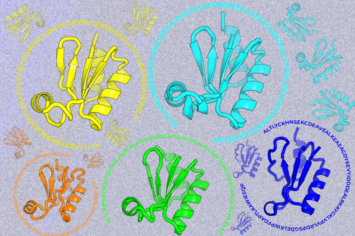
A new machine-learning framework aims to improve the success rate of computational protein design while moving away from results that reproduce sequences found in nature.