Amyloid fibrils are implicated in a myriad of human diseases. A striking observation is that fibrils extracted from diseased tissues are characterized by a restricted set of folds unique to the specific pathology. In contrast, fibrils grown in vitro exhibit extensive structural diversity, suggesting that specific environmental and biochemical mechanisms in vivo enforce structural selectivity. Here, we combine two-dimensional infrared (2D IR) spectroscopy and cryo-electron microscopy (cryo-EM) to investigate the mechanisms governing polymorph formation in the human Islet Amyloid Polypeptide (hIAPP). We demonstrate that 2D IR can resolve populations of distinct polymorphs identified by cryo-EM, enabling rapid label-free screening of conditions prior to labor-intensive microscopy screening. We find that conditions favoring secondary nucleation, such as high protein concentration, increase polymorphic diversity. Crucially, cryo-EM reveals that formed by secondary nucleation do not structurally replicate the parent template. Finally, by selectively inhibiting secondary nucleation using the C-terminal domain of the DNAJB6 chaperone, we steer aggregation toward a monomorphic state. These findings highlight the critical role of molecular chaperones in fibril polymorph selection.
Cryo-electron microscopy (cryo-EM) studies of amyloid fibrils have revealed endpoint structures of disease-relevant filaments and polymorphic intermediates formed during in vitro assembly of prion-like proteins. However, how transmissible prion or prion-like amyloids evolve during de novo formation and maturation in living cells remains unknown. Here, using the yeast prion [PSI+] as a model, we isolated Sup35NM amyloid fibrils from successive stages of [PSI+] maturation in Saccharomyces cerevisiae and characterised their near-atomic structures and population-level structural diversity by combining cryo-EM and atomic force microscopy. We show that intermediate and mature states differ in predominant fibril structure and the regions of the Sup35 sequence incorporated into the core, and that structural diversity decreases during maturation. Curing of [PSI+] at the mature state by guanidine hydrochloride (GdnHCl), which selectively inhibits ATPase activity of the chaperone Hsp104, restored both the predominant intermediate amyloid structure and the broader structural diversity characteristic of the intermediate state. In addition, Hsp104, Ssa1 (Hsp70) and Sis1 (Hsp40) associate differently with fibrils from the two states. Together, these findings provide direct structural evidence for amyloid evolution in vivo and support a chaperone-mediated mechanism of conformer selection within a polymorphic amyloid population.
Ziang Wang, Samantha L. Weetman, Barbara Altenhuber et al.· bioRxiv· 0 citations
Amyloid polymorphism is important in neurodegenerative and metabolic disease. Understanding fibril molecular polymorphism is critical to understanding why certain protein aggregates made from the same protein are pathogenic while others are benign. Here, we generate two polymorphs of human insulin, as a model system, induced by a thermal change in the nucleation (lag) phase. Both amyloid strains predominantly exhibit β-sheet structures; however, the cold-induced fibrils (formed under a two-h cold (4 °C) shock) display a more heterogeneous structure compared to the more uniform content in the conventional warm fibrils (formed at 37 °C the whole time). These structural variations highlight the complexity of the nucleation and growth mechanisms, with cold fibrils potentially trapped in non-equilibrium states due to a higher nucleation barrier. We find that the functional activity of cold/warm fibrils is unique in in vitro seeding/amyloid propagation, stability against inhibition, and cellular cytotoxicity. Our results show how amyloid polymorphs could differentially modulate pathogenicity. Moreover, the importance of environmental conditions on the molecular features of amyloid nucleation may be critical for design and stability of future protein biologics beyond therapeutic insulin.
Elnaz Hosseini, Saeid Hadi Alijanvand, Y. Kan et al.· Communications Chemistry· 0 citations
Alpha-synuclein is an intensely studied intrinsically disordered protein whose aggregation into amyloid fibrils is connected to the progression of several neurodegenerative diseases, most commonly Parkinson’s Disease. A remarkable feature that has emerged from this research is how easy it is to induce the protein to aggregate in vitro into a wide range of amyloid fibrils that appear to resemble the aggregates found in Lewy bodies in diseases like Parkinson’s while at the same time how difficult it is to produce aggregates whose fold truly represents the disease-associated amyloids at the atomic level. In an effort to produce the disease-relevant fibrils in vitro we have analyzed over 60 independent samples by cryo-electron microscopy using helical reconstruction to obtain atomic resolution models for most of the samples. While not yet achieving our original goal, we have found that several overlooked parameters influence the structural outcomes of alpha-synuclein aggregation, in particular protein purity, preparation of the monomeric starting material and agitation method.
Lukas Frey, D. Rhyner, Witek Kwiatkowski et al.· bioRxiv· 1 citation
Here, we present a comprehensive, topology-driven analysis of 543 amyloid structures (all cryo-EM-determined parallel amyloid fibril structures available in the PDB and the Amyloid Atlas, covering multiple protein types, notably Tau and α-synuclein) utilizing a novel automated protocol, ACWF. By calculating per-residue interface descriptors─shape complementarity (Sc), buried surface area (Ab), and surface detail index (SDi)─via a sliding window approach, we quantify structural packing and interdigitation across ∼30,000 local interfaces. We introduce a sequence-overlap-modified RMSD (RMSDmod) metric for hierarchical clustering to robustly classify polymorphs and quantify structural diversity. Our results reveal that mature amyloid fibrils contain a mixture of tightly and loosely packed regions, with distinct interaction hot-spots characteristic to polymorph families. Clustering successfully distinguishes disease-specific topologies and tracks maturation pathways, demonstrating that ex vivo fibrils rearrange to more compact structures indicated by more buried side chains (larger Ab) and typically lowered Sc values compared to that seen in the case of in vitro produced fibrils formed over shorter time scales. This work establishes a topology- and interface-based framework that links residue-level interactions to polymorphic fibril evolution, exploring the most compacted as well as accessible and attackable regions.
M. Sulyok-Eiler, V. Harmat, András Perczel· Journal of Chemical Informat...· 0 citations
Related blog posts
MIT News · Artificial Intelligence· news.mit.eduAug 27, 2026
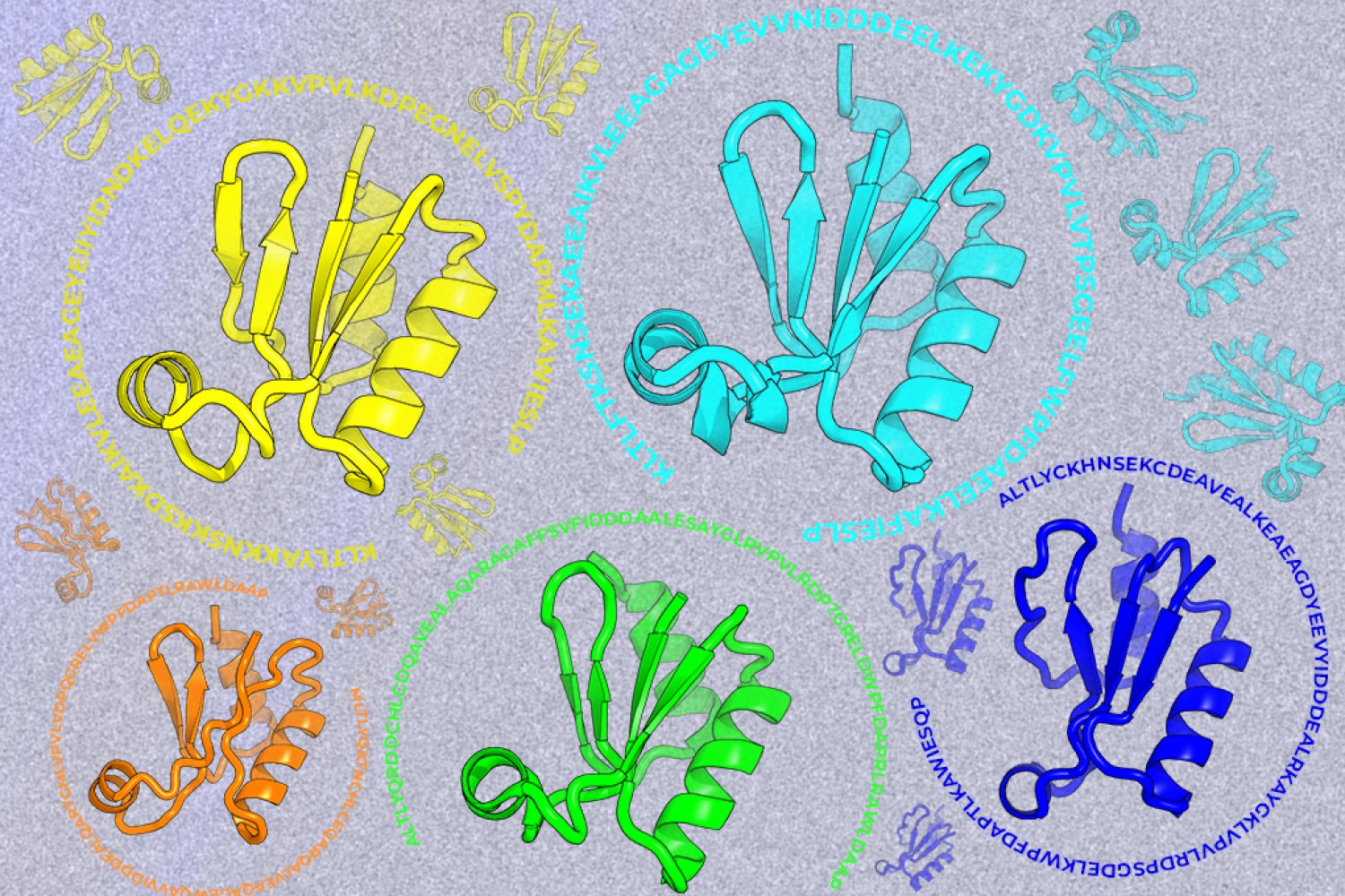
A new machine-learning framework aims to improve the success rate of computational protein design while moving away from results that reproduce sequences found in nature.