Skip to content
#protein folding Open access

So similar, yet so different: α-synuclein recognition of the isoform-specific VDAC protein–lipid interface

Oct 2026 · bioRxiv · 0 citations
Biology

TL;DR

Comparative analysis of α-synuclein interaction with three mammalian VDAC isoforms, complemented by corresponding data for VDAC2 substates and results from molecular dynamics simulations, reveals a new step in the chain of events leading to VDAC blockage.

Abstract

In mammals, VDAC exists as three isoforms – VDAC1, VDAC2, and VDAC3 – each characterized by a distinct tissue-dependent distribution and physiologic role. Isoform-specific knockout studies in cells and mice demonstrate distinct physiological roles for each VDAC isoform. The isoforms share about 70% sequence homology and a conserved β-barrel structure and, when reconstituted into planar lipid membranes, form similar highly conductive, anion-selective, voltage-gated channels. A natural question, then, is how relatively minor differences in isoform sequences lead to striking differences in their interactions with protein partners and, consequently, in their physiological functions. To address this question, we used a small, intrinsically disordered cytosolic protein α-synuclein (αSyn) as a sensitive molecular probe. A comparative study with recombinant VDACs reconstituted into planar lipid membranes shows that, depending on the isoform, the αSyn-induced blockage exhibits up to 100-fold differences in on-rates and equilibrium constants. The isoforms also display distinct translocation regimes for αSyn, as evidenced by altered dependences of the off-rates on the applied voltage. Deletion of cysteine residues has a significant effect on αSyn binding. Combined with corresponding data obtained for VDAC2 substates – which show an order of magnitude variation in on-rates while maintaining the conserved equilibrium constants – and the results of molecular dynamics simulations, these findings point to a previously unrecognized step in VDAC inhibition by αSyn: an isoform-specific αSyn interaction with the VDAC protein-lipid interface. This finding opens a new paradigm for understanding VDAC isoform functions and developing isoform-specific therapeutics. New & Noteworthy Comparative analysis of α-synuclein interaction with three mammalian VDAC isoforms, complemented by corresponding data for VDAC2 substates and results from molecular dynamics simulations, reveals a new step in the chain of events leading to VDAC blockage. Specifically, our findings point to the isoform-specific interaction of αSyn with VDAC protein-lipid interface, thereby guiding a new strategy in the search for VDAC-targeting drugs.
Read PDF

Similar papers

Open access Oct 2026

Towards VDAC pharmacology: Developing small-peptide channel inhibitors inspired by nature

The Voltage-Dependent Anion Channel (VDAC) forms a unique pathway across the mitochondrial outer membrane for small ions and water-soluble metabolites, such as ATP and ADP. Despite VDAC’s involvement in both normal regulation of respiration and in a wide variety of mitochondria-associated pathologies, there are, surpri...

T. Rostovtseva, P. Gurnev, D. Hoogerheide et al. · 0 citations
Open access Sep 2026

Phosphorylation of α-Synuclein Directs Trafficking of the v-SNARE Synaptobrevin-2

α-Synuclein has been extensively studied for its role in Parkinson's disease; however, its native function remains unclear. Several lines of evidence suggest it modulates presynaptic function. As many presynaptic proteins have their functions regulated by phosphorylation, we sought to determine whether α-synuclein, a k...

Elyas H. Arvell, James P. Carroll, Holly Melland et al. · 0 citations
Sep 2026

Electrostatic Regulation of Alpha-Synuclein Membrane Binding Couples Conformational Release to Aggregation Kinetics

These results identify a membrane charge density of 25–35% anionic lipid range as an electrostatic transition region in which the aggregation-prone NAC domain becomes solvent-exposed while the N-terminal anchor remains membrane-associated.

Mahdi Lavasani, Ping Lu, Shehani V. Siripala et al. · 0 citations
#protein folding Review Open access Oct 2026

Investigating protein conformational ensembles by native mass spectrometry and complementary biophysical techniques—the case of α-synuclein

One of the challenges of modern structural biology is to account for protein structural dynamics. Intrinsically disordered proteins (IDPs), which do not fold spontaneously into a defined conformation, existing, instead, as highly heterogeneous conformational ensembles, provide an extreme and challenging example of prot...

S. Brocca, E. Ponzini, C. Santambrogio et al. · 0 citations
Sep 2026

AQP4 Extended Region Binds to α-Syntrophin with a Novel Binding Motif.

Aquaporin-4 (AQP4) is an integral membrane protein channel specific for water molecules. It is highly polarized in the endfeet of astrocytes in the central nervous system where it allows water-ion homeostasis. AQP4 has two N-terminal isoforms: M1 (long) and M23 (short). Both isoforms retain their function but differ in...

Eric P. Jacobo, Michael J. Martinez, Barbara Barile et al. · 0 citations

Related blog posts

Google DeepMind Blog Sep 30, 2026

Introducing SynthID Bio

Proof of concept for watermarking AI-generated proteins while preserving biological function.

MIT News · Artificial Intelligence Aug 27, 2026

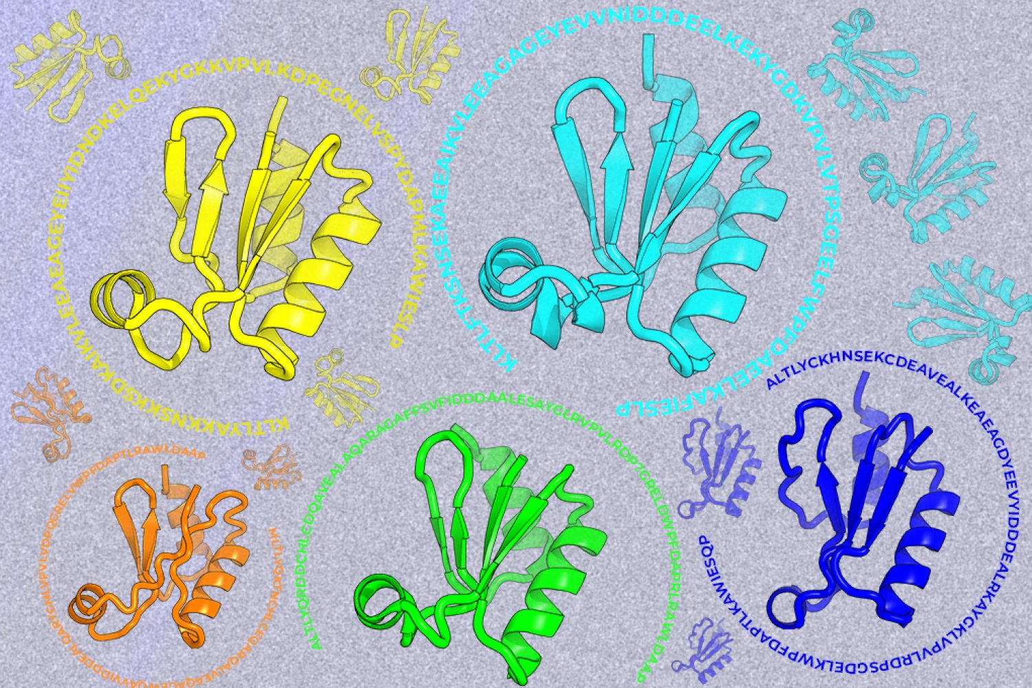
Looking beyond natural sequences

A new machine-learning framework aims to improve the success rate of computational protein design while moving away from results that reproduce sequences found in nature.

We use cookies to run the site and, with your consent, for analytics and to show ads. See our Cookie Policy.